1SDW

Reduced (Cu+) peptidylglycine alpha-hydroxylating monooxygenase with bound peptide and dioxygen


Experimental Data Snapshot

  • Method: X-RAY DIFFRACTION
  • Resolution: 1.85 Å
  • R-Value Free: 0.226 
  • R-Value Work: 0.197 
  • R-Value Observed: 0.197 

wwPDB Validation   3D Report Full Report


This is version 1.3 of the entry. See complete history


Literature

Dioxygen binds end-on to mononuclear copper in a precatalytic enzyme complex.

Prigge, S.T.Eipper, B.A.Mains, R.E.Amzel, L.M.

(2004) Science 304: 864-867

  • DOI: https://doi.org/10.1126/science.1094583
  • Primary Citation of Related Structures:  
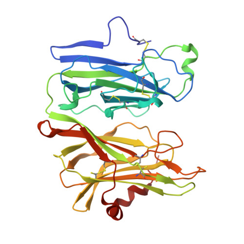
    1SDW

  • PubMed Abstract: 

    Copper active sites play a major role in enzymatic activation of dioxygen. We trapped the copper-dioxygen complex in the enzyme peptidylglycine-alphahydroxylating monooxygenase (PHM) by freezing protein crystals that had been soaked with a slow substrate and ascorbate in the presence of oxygen. The x-ray crystal structure of this precatalytic complex, determined to 1.85-angstrom resolution, shows that oxygen binds to one of the coppers in the enzyme with an end-on geometry. Given this structure, it is likely that dioxygen is directly involved in the electron transfer and hydrogen abstraction steps of the PHM reaction. These insights may apply to other copper oxygen-activating enzymes, such as dopamine beta-monooxygenase, and to the design of biomimetic complexes.


  • Organizational Affiliation

    Department of Microbiology and Molecular Immunology, The Bloomberg School of Public Health, Johns Hopkins University, Baltimore, MD, USA.


Macromolecules
Find similar proteins by:  (by identity cutoff)  |  3D Structure
Entity ID: 1
MoleculeChains Sequence LengthOrganismDetailsImage
Peptidyl-glycine alpha-amidating monooxygenase314Rattus norvegicusMutation(s): 0 
Gene Names: PAM
EC: 1.14.17.3
UniProt
Find proteins for P14925 (Rattus norvegicus)
Explore P14925 
Go to UniProtKB:  P14925
Entity Groups  
Sequence Clusters30% Identity50% Identity70% Identity90% Identity95% Identity100% Identity
UniProt GroupP14925
Sequence Annotations
Expand
  • Reference Sequence
Small Molecules
Ligands 5 Unique
IDChains Name / Formula / InChI Key2D Diagram3D Interactions
IYT
Query on IYT

Download Ideal Coordinates CCD File 
F [auth A],
G [auth A],
H [auth A]
N-ALPHA-ACETYL-3,5-DIIODOTYROSYL-D-THREONINE
C15 H18 I2 N2 O6
TWMKRGDZEJLDDH-LKXWSVAYSA-N
GOL
Query on GOL

Download Ideal Coordinates CCD File 
I [auth A]
J [auth A]
K [auth A]
L [auth A]
M [auth A]
I [auth A],
J [auth A],
K [auth A],
L [auth A],
M [auth A],
N [auth A]
GLYCEROL
C3 H8 O3
PEDCQBHIVMGVHV-UHFFFAOYSA-N
CU
Query on CU

Download Ideal Coordinates CCD File 
B [auth A],
C [auth A]
COPPER (II) ION
Cu
JPVYNHNXODAKFH-UHFFFAOYSA-N
NI
Query on NI

Download Ideal Coordinates CCD File 
D [auth A]NICKEL (II) ION
Ni
VEQPNABPJHWNSG-UHFFFAOYSA-N
OXY
Query on OXY

Download Ideal Coordinates CCD File 
E [auth A]OXYGEN MOLECULE
O2
MYMOFIZGZYHOMD-UHFFFAOYSA-N
Experimental Data & Validation

Experimental Data

  • Method: X-RAY DIFFRACTION
  • Resolution: 1.85 Å
  • R-Value Free: 0.226 
  • R-Value Work: 0.197 
  • R-Value Observed: 0.197 
  • Space Group: P 21 21 21
Unit Cell:
Length ( Å )Angle ( ˚ )
a = 69.478α = 90
b = 68.979β = 90
c = 82.108γ = 90
Software Package:
Software NamePurpose
DENZOdata reduction
SCALEPACKdata scaling
CNSrefinement
CNSphasing

Structure Validation

View Full Validation Report


Entry History 

Deposition Data

Revision History  (Full details and data files)

  • Version 1.0: 2004-05-11
    Type: Initial release
  • Version 1.1: 2008-04-29
    Changes: Version format compliance
  • Version 1.2: 2011-07-13
    Changes: Non-polymer description, Version format compliance
  • Version 1.3: 2023-08-23
    Changes: Data collection, Database references, Derived calculations, Refinement description