1SDD

Crystal Structure of Bovine Factor Vai


Experimental Data Snapshot

  • Method: X-RAY DIFFRACTION
  • Resolution: 2.80 Å
  • R-Value Free: 0.296 
  • R-Value Work: 0.252 
  • R-Value Observed: 0.262 

wwPDB Validation   3D Report Full Report


Ligand Structure Quality Assessment 


This is version 2.0 of the entry. See complete history


Literature

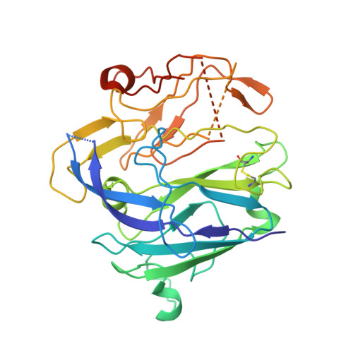
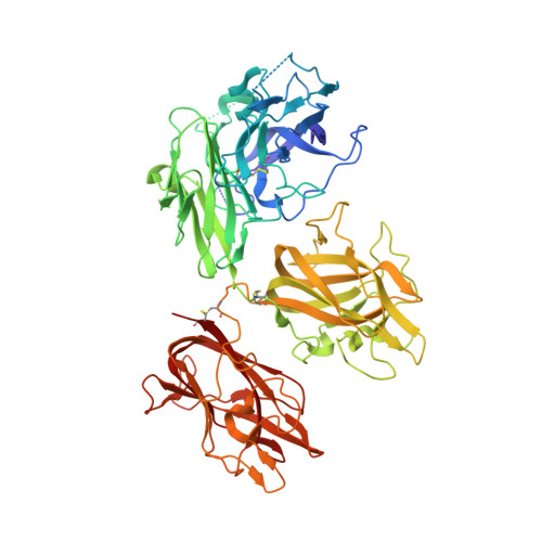
The crystal structure of activated protein C-inactivated bovine factor Va: Implications for cofactor function.

Adams, T.E.Hockin, M.F.Mann, K.G.Everse, S.J.

(2004) Proc Natl Acad Sci U S A 101: 8918-8923

  • DOI: https://doi.org/10.1073/pnas.0403072101
  • Primary Citation of Related Structures:  
    1SDD

  • PubMed Abstract: 

    In vertebrate hemostasis, factor Va serves as the cofactor in the prothrombinase complex that results in a 300,000-fold increase in the rate of thrombin generation compared with factor Xa alone. Structurally, little is known about the mechanism by which factor Va alters catalysis within this complex. Here, we report a crystal structure of protein C inactivated factor Va (A1.A3-C1-C2) that depicts a previously uncharacterized domain arrangement. This orientation has implications for binding to membranes essential for function. A high-affinity calcium-binding site and a copper-binding site have both been identified. Surprisingly, neither shows a direct involvement in chain association. This structure represents the largest physiologically relevant fragment of factor Va solved to date and provides a new scaffold for the future generation of models of coagulation cofactors.


  • Organizational Affiliation

    Department of Biochemistry, College of Medicine, University of Vermont, 89 Beaumont Avenue, Burlington, VT 05405, USA.


Macromolecules
Find similar proteins by:  (by identity cutoff)  |  3D Structure
Entity ID: 1
MoleculeChains Sequence LengthOrganismDetailsImage
Coagulation factor V306Bos taurusMutation(s): 0 
Membrane Entity: Yes 
UniProt
Find proteins for Q28107 (Bos taurus)
Explore Q28107 
Go to UniProtKB:  Q28107
Entity Groups  
Sequence Clusters30% Identity50% Identity70% Identity90% Identity95% Identity100% Identity
UniProt GroupQ28107
Sequence Annotations
Expand
  • Reference Sequence
Find similar proteins by:  (by identity cutoff)  |  3D Structure
Entity ID: 2
MoleculeChains Sequence LengthOrganismDetailsImage
Coagulation factor V647Bos taurusMutation(s): 0 
Membrane Entity: Yes 
UniProt
Find proteins for Q28107 (Bos taurus)
Explore Q28107 
Go to UniProtKB:  Q28107
Entity Groups  
Sequence Clusters30% Identity50% Identity70% Identity90% Identity95% Identity100% Identity
UniProt GroupQ28107
Sequence Annotations
Expand
  • Reference Sequence
Experimental Data & Validation

Experimental Data

  • Method: X-RAY DIFFRACTION
  • Resolution: 2.80 Å
  • R-Value Free: 0.296 
  • R-Value Work: 0.252 
  • R-Value Observed: 0.262 
  • Space Group: P 21 21 21
Unit Cell:
Length ( Å )Angle ( ˚ )
a = 63.37α = 90
b = 86.56β = 90
c = 229.2γ = 90
Software Package:
Software NamePurpose
DENZOdata reduction
SCALEPACKdata scaling
MLPHAREphasing
CNSrefinement

Structure Validation

View Full Validation Report


Ligand Structure Quality Assessment 


Entry History 

Deposition Data

Revision History  (Full details and data files)

  • Version 1.0: 2004-06-29
    Type: Initial release
  • Version 1.1: 2007-10-16
    Changes: Version format compliance
  • Version 1.2: 2011-07-13
    Changes: Derived calculations, Version format compliance
  • Version 2.0: 2020-07-29
    Type: Remediation
    Reason: Carbohydrate remediation
    Changes: Advisory, Atomic model, Data collection, Derived calculations, Non-polymer description, Structure summary