1S47

Crystal structure analysis of the DNA quadruplex d(TGGGGT)S2


Experimental Data Snapshot

  • Method: X-RAY DIFFRACTION
  • Resolution: 2.50 Å
  • R-Value Free: 0.311 
  • R-Value Work: 0.224 
  • R-Value Observed: 0.224 

wwPDB Validation   3D Report Full Report


This is version 1.3 of the entry. See complete history


Literature

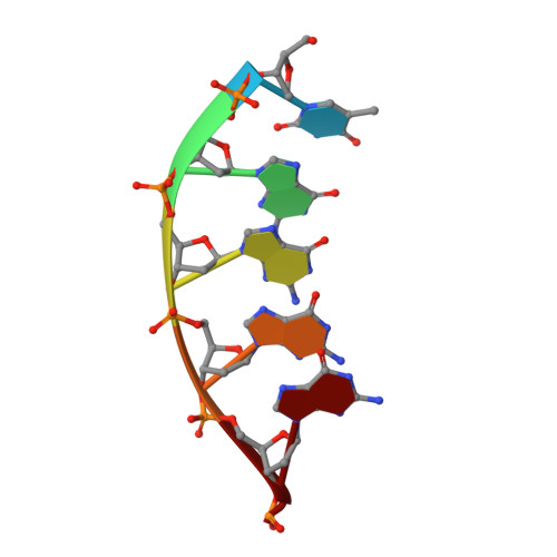
A Thymine tetrad in d(TGGGGT) quadruplexes stabilized with Tl+1/Na+1 ions

Caceres, C.Wright, G.Gouyette, C.Parkinson, G.Subirana, J.A.

(2004) Nucleic Acids Res 32: 1097-1102

  • DOI: https://doi.org/10.1093/nar/gkh269
  • Primary Citation of Related Structures:  
    1S45, 1S47

  • PubMed Abstract: 

    We report two new structures of the quadruplex d(TGGGGT)4 obtained by single crystal X-ray diffraction. In one of them a thymine tetrad is found. Thus the yeast telomere sequences d(TG1-3) might be able to form continuous quadruplex structures, involving both guanine and thymine tetrads. Our study also shows substantial differences in the arrangement of thymines when compared with previous studies. We find five different types of organization: (i) groove binding with hydrogen bonds to guanines from a neighbour quadruplex; (ii) partially ordered groove binding, without any hydrogen bond; (iii) stacked thymine triads, formed at the 3'ends of the quadruplexes; (iv) a thymine tetrad between two guanine tetrads. Thymines are stabilized in pairs by single hydrogen bonds. A central sodium ion interacts with two thymines and contributes to the tetrad structure. (v) Completely disordered thymines which do not show any clear location in the crystal. The tetrads are stabilized by either Na+ or Tl+ ions. We show that by using MAD methods, Tl+ can be unambiguously located and distinguished from Na+. We can thus determine the preference for either ion in each ionic site of the structure under the conditions used by us.


  • Organizational Affiliation

    Departament d'Enginyeria Química, Universitat Politècnica de Catalunya, Avinguda Diagonal 647, E-08028 Barcelona, Spain.


Macromolecules

Find similar nucleic acids by:  Sequence   |   3D Structure  

Entity ID: 1
MoleculeChains LengthOrganismImage
5'-D(*TP*GP*GP*GP*GP*T)-3'
A, B, C, D, E
A, B, C, D, E, F, G, H, I, J, K, L
6N/A
Sequence Annotations
Expand
  • Reference Sequence
Small Molecules
Ligands 2 Unique
IDChains Name / Formula / InChI Key2D Diagram3D Interactions
TL
Query on TL

Download Ideal Coordinates CCD File 
O [auth B]
P [auth D]
S [auth E]
V [auth I]
W [auth I]
O [auth B],
P [auth D],
S [auth E],
V [auth I],
W [auth I],
X [auth I]
THALLIUM (I) ION
Tl
ZLUSCZLCHQSJRU-UHFFFAOYSA-N
NA
Query on NA

Download Ideal Coordinates CCD File 
M [auth A]
N [auth A]
Q [auth E]
R [auth E]
T [auth F]
M [auth A],
N [auth A],
Q [auth E],
R [auth E],
T [auth F],
U [auth H],
Y [auth K]
SODIUM ION
Na
FKNQFGJONOIPTF-UHFFFAOYSA-N
Experimental Data & Validation

Experimental Data

  • Method: X-RAY DIFFRACTION
  • Resolution: 2.50 Å
  • R-Value Free: 0.311 
  • R-Value Work: 0.224 
  • R-Value Observed: 0.224 
  • Space Group: P 1 21 1
Unit Cell:
Length ( Å )Angle ( ˚ )
a = 28.319α = 90
b = 56.341β = 99.17
c = 51.746γ = 90
Software Package:
Software NamePurpose
CNSrefinement
DENZOdata reduction
SCALEPACKdata scaling
EPMRphasing

Structure Validation

View Full Validation Report


Entry History 

Deposition Data

Revision History  (Full details and data files)

  • Version 1.0: 2004-02-24
    Type: Initial release
  • Version 1.1: 2008-04-29
    Changes: Version format compliance
  • Version 1.2: 2011-07-13
    Changes: Version format compliance
  • Version 1.3: 2023-08-23
    Changes: Data collection, Database references, Derived calculations, Refinement description