1RTA

CRYSTAL STRUCTURE DISPOSITION OF THYMIDYLIC ACID TETRAMER IN COMPLEX WITH RIBONUCLEASE A


Experimental Data Snapshot

  • Method: X-RAY DIFFRACTION
  • Resolution: 2.50 Å
  • R-Value Work: 0.235 
  • R-Value Observed: 0.235 

wwPDB Validation   3D Report Full Report


This is version 1.2 of the entry. See complete history


Literature

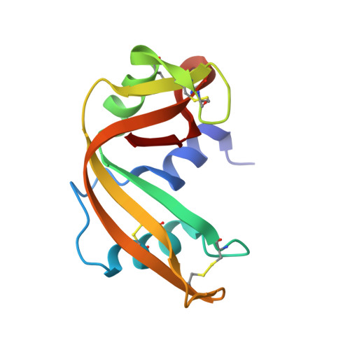
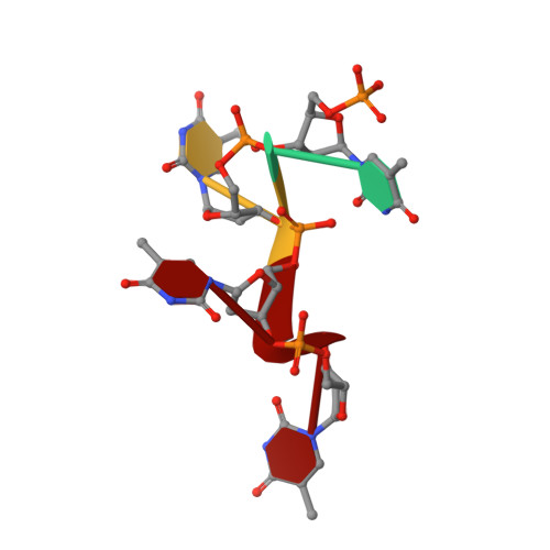
Crystal structure disposition of thymidylic acid tetramer in complex with ribonuclease A.

Birdsall, D.L.McPherson, A.

(1992) J Biol Chem 267: 22230-22236

  • Primary Citation of Related Structures:  
    1RTA, 1RTB

  • PubMed Abstract: 

    The crystal structure of ribonuclease A with bound thymidylic acid tetramer is reported at 2.5-A resolution. The diffusion of the tetramer into native orthorhombic crystals of the ribonuclease allows for the formation of a structurally stable complex where the single-stranded nucleic acid enters and leaves the enzyme's catalytic region in a persistent 5'-3' direction. The binding of the tetramer to the enzyme's surface is facilitated and mediated by electrostatic interactions between basic protein residues and nucleotide phosphates. Two pyrimidine nucleotides are bound to the enzyme's active site in a manner similar to that observed for other complexes between ribonuclease A and nucleic acid oligomers.


  • Organizational Affiliation

    Division of Biomedical Sciences, Lawrence Livermore National Laboratory, University of California, Livermore 94550.


Macromolecules

Find similar proteins by:  (by identity cutoff)  |  3D Structure
Entity ID: 2
MoleculeChains Sequence LengthOrganismDetailsImage
PROTEIN (RIBONUCLEASE A (E.C.3.1.27.5))B [auth E]124Bos taurusMutation(s): 0 
EC: 3.1.27.5
UniProt
Find proteins for P61823 (Bos taurus)
Explore P61823 
Go to UniProtKB:  P61823
Entity Groups  
Sequence Clusters30% Identity50% Identity70% Identity90% Identity95% Identity100% Identity
UniProt GroupP61823
Sequence Annotations
Expand
  • Reference Sequence

Find similar nucleic acids by:  Sequence   |   3D Structure  

Entity ID: 1
MoleculeChains LengthOrganismImage
DNA (5'-D(*TP*TP*TP*T)-3')A [auth C]4N/A
Sequence Annotations
Expand
  • Reference Sequence
Experimental Data & Validation

Experimental Data

  • Method: X-RAY DIFFRACTION
  • Resolution: 2.50 Å
  • R-Value Work: 0.235 
  • R-Value Observed: 0.235 
  • Space Group: P 21 21 21
Unit Cell:
Length ( Å )Angle ( ˚ )
a = 44.9α = 90
b = 75β = 90
c = 43.6γ = 90
Software Package:
Software NamePurpose
X-PLORrefinement
CORELSrefinement

Structure Validation

View Full Validation Report


Entry History 

Deposition Data

Revision History  (Full details and data files)

  • Version 1.0: 1993-10-31
    Type: Initial release
  • Version 1.1: 2008-05-22
    Changes: Version format compliance
  • Version 1.2: 2011-07-13
    Changes: Version format compliance