1R30

The Crystal Structure of Biotin Synthase, an S-Adenosylmethionine-Dependent Radical Enzyme


Experimental Data Snapshot

  • Method: X-RAY DIFFRACTION
  • Resolution: 3.40 Å
  • R-Value Free: 0.300 
  • R-Value Work: 0.256 
  • R-Value Observed: 0.256 

wwPDB Validation   3D Report Full Report


This is version 1.4 of the entry. See complete history


Literature

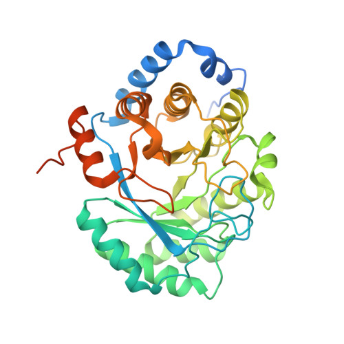
Crystal structure of biotin synthase, an S-adenosylmethionine-dependent radical enzyme.

Berkovitch, F.Nicolet, Y.Wan, J.T.Jarrett, J.T.Drennan, C.L.

(2004) Science 303: 76-79

  • DOI: https://doi.org/10.1126/science.1088493
  • Primary Citation of Related Structures:  
    1R30

  • PubMed Abstract: 

    The crystal structure of biotin synthase from Escherichia coli in complex with S-adenosyl-L-methionine and dethiobiotin has been determined to 3.4 angstrom resolution. This structure addresses how "AdoMet radical" or "radical SAM" enzymes use Fe4S4 clusters and S-adenosyl-L-methionine to generate organic radicals. Biotin synthase catalyzes the radical-mediated insertion of sulfur into dethiobiotin to form biotin. The structure places the substrates between the Fe4S4 cluster, essential for radical generation, and the Fe2S2 cluster, postulated to be the source of sulfur, with both clusters in unprecedented coordination environments.


  • Organizational Affiliation

    Department of Chemistry, Massachusetts Institute of Technology, Cambridge, MA 02139, USA.


Macromolecules
Find similar proteins by:  (by identity cutoff)  |  3D Structure
Entity ID: 1
MoleculeChains Sequence LengthOrganismDetailsImage
Biotin synthase
A, B
369Escherichia coliMutation(s): 0 
Gene Names: BIOB
EC: 2.8.1.6
UniProt
Find proteins for P12996 (Escherichia coli (strain K12))
Explore P12996 
Go to UniProtKB:  P12996
Entity Groups  
Sequence Clusters30% Identity50% Identity70% Identity90% Identity95% Identity100% Identity
UniProt GroupP12996
Sequence Annotations
Expand
  • Reference Sequence
Small Molecules
Ligands 5 Unique
IDChains Name / Formula / InChI Key2D Diagram3D Interactions
SAM
Query on SAM

Download Ideal Coordinates CCD File 
C [auth A],
H [auth B]
S-ADENOSYLMETHIONINE
C15 H22 N6 O5 S
MEFKEPWMEQBLKI-FCKMPRQPSA-N
SF4
Query on SF4

Download Ideal Coordinates CCD File 
D [auth A],
I [auth B]
IRON/SULFUR CLUSTER
Fe4 S4
LJBDFODJNLIPKO-UHFFFAOYSA-N
DTB
Query on DTB

Download Ideal Coordinates CCD File 
F [auth A],
K [auth B]
6-(5-METHYL-2-OXO-IMIDAZOLIDIN-4-YL)-HEXANOIC ACID
C10 H18 N2 O3
AUTOLBMXDDTRRT-JGVFFNPUSA-N
FES
Query on FES

Download Ideal Coordinates CCD File 
E [auth A],
J [auth B]
FE2/S2 (INORGANIC) CLUSTER
Fe2 S2
NIXDOXVAJZFRNF-UHFFFAOYSA-N
TRS
Query on TRS

Download Ideal Coordinates CCD File 
G [auth A]2-AMINO-2-HYDROXYMETHYL-PROPANE-1,3-DIOL
C4 H12 N O3
LENZDBCJOHFCAS-UHFFFAOYSA-O
Experimental Data & Validation

Experimental Data

  • Method: X-RAY DIFFRACTION
  • Resolution: 3.40 Å
  • R-Value Free: 0.300 
  • R-Value Work: 0.256 
  • R-Value Observed: 0.256 
  • Space Group: P 31 2 1
Unit Cell:
Length ( Å )Angle ( ˚ )
a = 155.69α = 90
b = 155.69β = 90
c = 90.88γ = 120
Software Package:
Software NamePurpose
CNSrefinement
CBASSdata collection
Adxvdata processing
XDSdata scaling
SOLVEphasing

Structure Validation

View Full Validation Report


Entry History 

Deposition Data

Revision History  (Full details and data files)

  • Version 1.0: 2004-01-13
    Type: Initial release
  • Version 1.1: 2008-04-29
    Changes: Version format compliance
  • Version 1.2: 2011-07-13
    Changes: Version format compliance
  • Version 1.3: 2017-10-11
    Changes: Refinement description
  • Version 1.4: 2024-02-14
    Changes: Data collection, Database references, Derived calculations