1QTF

CRYSTAL STRUCTURE OF EXFOLIATIVE TOXIN B


Experimental Data Snapshot

  • Method: X-RAY DIFFRACTION
  • Resolution: 2.40 Å
  • R-Value Free: 0.290 
  • R-Value Work: 0.204 

wwPDB Validation   3D Report Full Report


This is version 1.4 of the entry. See complete history


Literature

The crystal structure of exfoliative toxin B: a superantigen with enzymatic activity.

Vath, G.M.Earhart, C.A.Monie, D.D.Iandolo, J.J.Schlievert, P.M.Ohlendorf, D.H.

(1999) Biochemistry 38: 10239-10246

  • DOI: https://doi.org/10.1021/bi990721e
  • Primary Citation of Related Structures:  
    1QTF

  • PubMed Abstract: 

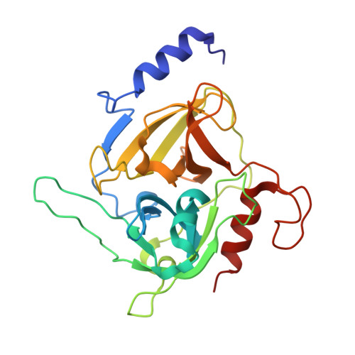
    The exfoliative toxins (ETs) cause staphylococcal scalded skin syndrome, a disease characterized by specific separation of layers of the skin. Evidence suggests that the toxins act as serine proteases, though the specific substrate and mode of action are not known for certain. The crystal structure of exfoliative toxin A (ETA) was reported earlier and shown to be similar to that of the chymotrypsin-like serine proteases. Here, we report the 2.4 A resolution crystal structure of the other exfoliative toxin, ETB, which is 40% identical to ETA. The overall structures of ETA and ETB are similar including the positions of key residues within the active site. The structure of ETB supports the previous findings that the ETs are serine proteases that cleave substrates after glutamic acid residues. In this study we also discuss a number of structural differences including a large 14 residue loop insertion which may be a key feature involved in the differing biological properties of the ETs, particularly the pyrogenic and lethal activities of ETB not shared by ETA.


  • Organizational Affiliation

    Department of Biochemistry, Molecular Biology, and Biophysics, University of Minnesota Medical School, Minneapolis 55455, USA.


Macromolecules
Find similar proteins by:  (by identity cutoff)  |  3D Structure
Entity ID: 1
MoleculeChains Sequence LengthOrganismDetailsImage
EXFOLIATIVE TOXIN B246Staphylococcus aureusMutation(s): 0 
EC: 3.4.21
Membrane Entity: Yes 
UniProt
Find proteins for P09332 (Staphylococcus aureus)
Explore P09332 
Go to UniProtKB:  P09332
Entity Groups  
Sequence Clusters30% Identity50% Identity70% Identity90% Identity95% Identity100% Identity
UniProt GroupP09332
Sequence Annotations
Expand
  • Reference Sequence
Experimental Data & Validation

Experimental Data

  • Method: X-RAY DIFFRACTION
  • Resolution: 2.40 Å
  • R-Value Free: 0.290 
  • R-Value Work: 0.204 
  • Space Group: C 2 2 21
Unit Cell:
Length ( Å )Angle ( ˚ )
a = 69.97α = 90
b = 71.09β = 90
c = 108.37γ = 90
Software Package:
Software NamePurpose
X-PLORmodel building
X-PLORrefinement
X-GENdata reduction
X-GENdata scaling
X-PLORphasing

Structure Validation

View Full Validation Report


Entry History 

Deposition Data

Revision History  (Full details and data files)

  • Version 1.0: 1999-08-31
    Type: Initial release
  • Version 1.1: 2008-04-27
    Changes: Version format compliance
  • Version 1.2: 2011-07-13
    Changes: Version format compliance
  • Version 1.3: 2017-10-04
    Changes: Refinement description
  • Version 1.4: 2024-02-14
    Changes: Data collection, Database references