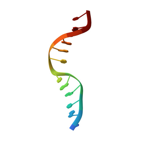
Solution structure of a five-adenine bulge loop within a DNA duplex.
Dornberger, U., Hillisch, A., Gollmick, F.A., Fritzsche, H., Diekmann, S.(1999) Biochemistry 38: 12860-12868
- PubMed: 10504256
- DOI: https://doi.org/10.1021/bi9906874
- Primary Citation of Related Structures:
1QSK - PubMed Abstract:
The three-dimensional solution structure of a DNA molecule of the sequence 5'-d(GCATCGAAAAAGCTACG)-3' paired with 5'-d(CGTAGCCGATGC)-3' containing a five-adenine bulge loop (dA(5)-bulge) between two double helical stems was determined by 2D (1)H and (31)P NMR, infrared, and Raman spectroscopy. The DNA in both stems adopt a classical B-form double helical structure with Watson-Crick base pairing and C2'-endo sugar conformation. In addition, the two dG/dC base pairs framing the dA(5)-bulge loop are formed and are stable at least up to 30 degrees C. The five adenine bases of the bulge loop are localized at intrahelical positions within the double helical stems. Stacking on the double helical stem is continued for the first four 5'-adenines in the bulge loop. The total rise (the height) of these four stacked adenines roughly equals the diameter of the double helical stem. The stacking interactions are broken between the last of these four 5'-adenines and the fifth loop adenine at the 3'-end. This 3'-adenine partially stacks on the other stem. The angle between the base planes of the two nonstacking adenines (A10 and A11) in the bulge loop reflects the kinking angle of the global DNA structure. The neighboring cytosines opposite the dA(5)-bulge (being parts of the bulge flanking base pairs) do not stack on one another. This disruption of stacking is characterized by a partial shearing of these bases, such that certain sequential NOEs for this base step are preserved. In the base step opposite the loop, an extraordinary hydrogen bond is observed between the phosphate backbone of the 5'-dC and the amino proton of the 3'-dC in about two-thirds of the conformers. This hydrogen bond probably contributes to stabilizing the global DNA structure. The dA(5)-bulge induces a local kink into the DNA molecule of about 73 degrees (+/-11 degrees ). This kinking angle and the mutual orientation of the two double helical stems agree well with results from fluorescence resonance energy transfer measurements of single- and double-bulge DNA molecules.
Organizational Affiliation:
Institut für Molekularbiologie, Friedrich-Schiller-Universität, Winzerlaer Strasse 10, D-07745 Jena and Institut für molekulare Biotechnologie, Beutenbergstrasse 11, D-07745 Jena, Germany.