1QPQ

Structure of Quinolinic Acid Phosphoribosyltransferase from Mycobacterium Tuberculosis: A Potential TB Drug Target


Experimental Data Snapshot

  • Method: X-RAY DIFFRACTION
  • Resolution: 2.45 Å
  • R-Value Free: 0.253 
  • R-Value Work: 0.178 

wwPDB Validation   3D Report Full Report


This is version 1.3 of the entry. See complete history


Literature

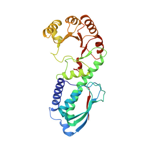
Crystal structure of quinolinic acid phosphoribosyltransferase from Mycobacterium tuberculosis: a potential TB drug target.

Sharma, V.Grubmeyer, C.Sacchettini, J.C.

(1998) Structure 6: 1587-1599

  • DOI: https://doi.org/10.1016/s0969-2126(98)00156-7
  • Primary Citation of Related Structures:  
    1QPN, 1QPO, 1QPQ, 1QPR

  • PubMed Abstract: 

    . Mycobacterium tuberculosis is the single most deadly human pathogen and is responsible for nearly three million deaths every year. Recent elucidation of the mode of action of isoniazid, a frontline antimycobacterial drug, suggests that NAD metabolism is extremely critical for this microorganism. M. tuberculosis depends solely on the de novo pathway to meet its NAD demand. Quinolinic acid phosphoribosyltransferase (QAPRTase), a key enzyme in the de novo biosynthesis of NAD, provides an attractive target for designing novel antitubercular drugs. . The X-ray crystal structure of the M. tuberculosis QAPRTase apoenzyme has been determined by multiple isomorphous replacement at 2.4 A resolution. Structures of the enzyme have also been solved in complex with the substrate quinolinic acid (QA), the inhibitory QA analog phthalic acid (PA), the product nicotinate mononucleotide (NAMN), and as a ternary complex with PA and a substrate analog, 5-phosphoribosyl-1-(beta-methylene)pyrophosphate (PRPCP). The structure of the nonproductive QAPRTase-PA-PRPCP Michaelis complex reveals a 5-phosphoribosyl-1-pyrophosphate-binding site that is different from the one observed in type I phosphoribosyltransferases (PRTases). The type II PRTase active site of QAPRTase undergoes conformational changes that appear to be important in determining substrate specificity and eliciting productive catalysis. . QAPRTase is the only known representative of the type II PRTase fold, an unusual alpha/beta barrel, and appears to represent convergent evolution for PRTase catalysis. The active site of type II PRTase bears little resemblance to the better known type I enzymes.


  • Organizational Affiliation

    Department of Biochemistry and Biophysics, Texas A&M University, College Station, TX 77843, USA.


Macromolecules
Find similar proteins by:  (by identity cutoff)  |  3D Structure
Entity ID: 1
MoleculeChains Sequence LengthOrganismDetailsImage
QUINOLINATE PHOSPHORIBOSYLTRANSFERASE
A, B, C, D, E
A, B, C, D, E, F
284Mycobacterium tuberculosis H37RvMutation(s): 0 
Gene Names: NADC
EC: 2.4.2.19
UniProt
Find proteins for P9WJJ7 (Mycobacterium tuberculosis (strain ATCC 25618 / H37Rv))
Explore P9WJJ7 
Go to UniProtKB:  P9WJJ7
Entity Groups  
Sequence Clusters30% Identity50% Identity70% Identity90% Identity95% Identity100% Identity
UniProt GroupP9WJJ7
Sequence Annotations
Expand
  • Reference Sequence
Small Molecules
Ligands 2 Unique
IDChains Name / Formula / InChI Key2D Diagram3D Interactions
NTM
Query on NTM

Download Ideal Coordinates CCD File 
H [auth A]
J [auth B]
L [auth C]
N [auth D]
P [auth E]
H [auth A],
J [auth B],
L [auth C],
N [auth D],
P [auth E],
R [auth F]
QUINOLINIC ACID
C7 H5 N O4
GJAWHXHKYYXBSV-UHFFFAOYSA-N
SO4
Query on SO4

Download Ideal Coordinates CCD File 
G [auth A]
I [auth B]
K [auth C]
M [auth D]
O [auth E]
G [auth A],
I [auth B],
K [auth C],
M [auth D],
O [auth E],
Q [auth F]
SULFATE ION
O4 S
QAOWNCQODCNURD-UHFFFAOYSA-L
Experimental Data & Validation

Experimental Data

  • Method: X-RAY DIFFRACTION
  • Resolution: 2.45 Å
  • R-Value Free: 0.253 
  • R-Value Work: 0.178 
  • Space Group: P 31
Unit Cell:
Length ( Å )Angle ( ˚ )
a = 100.34α = 90
b = 100.34β = 90
c = 145.658γ = 120
Software Package:
Software NamePurpose
X-PLORmodel building
X-PLORrefinement
DENZOdata reduction
SCALEPACKdata scaling
X-PLORphasing

Structure Validation

View Full Validation Report


Entry History 

Deposition Data

Revision History  (Full details and data files)

  • Version 1.0: 1998-11-25
    Type: Initial release
  • Version 1.1: 2007-10-21
    Changes: Version format compliance
  • Version 1.2: 2011-07-13
    Changes: Derived calculations, Version format compliance
  • Version 1.3: 2023-12-27
    Changes: Data collection, Database references, Derived calculations