1QMV

thioredoxin peroxidase B from red blood cells


Experimental Data Snapshot

  • Method: X-RAY DIFFRACTION
  • Resolution: 1.70 Å
  • R-Value Free: 0.256 
  • R-Value Work: 0.192 

wwPDB Validation   3D Report Full Report


This is version 1.4 of the entry. See complete history


Literature

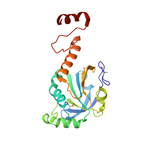
Crystal Structure of Decameric 2-Cys Peroxiredoxin from Human Erythrocytes at 1.7 A Resolution.

Schroder, E.Littlechild, J.A.Lebedev, A.A.Errington, N.Vagin, A.A.Isupov, M.N.

(2000) Structure 8: 605

  • DOI: https://doi.org/10.1016/s0969-2126(00)00147-7
  • Primary Citation of Related Structures:  
    1QMV

  • PubMed Abstract: 

    The peroxiredoxins (Prxs) are an emerging family of multifunctional enzymes that exhibit peroxidase activity in vitro, and in vivo participate in a range of cellular processes known to be sensitive to reactive oxygen species. Thioredoxin peroxidase B (TPx-B), a 2-Cys type II Prx from erythrocytes, promotes potassium efflux and down-regulates apoptosis and the recruitment of monocytes by endothelial tissue. The crystal structure of human decameric TPx-B purified from erythrocytes has been determined to 1.7 [corrected)] A resolution. The structure is a toroid comprising five dimers linked end-on through predominantly hydrophobic interactions, and is proposed to represent an intermediate in the in vivo reaction cycle. In the crystal structure, Cys51, the site of peroxide reduction, is oxidised to cysteine sulphinic acid. The residue Cys172, lies approximately 10 A away from Cys51 [corrected]. The oxidation of Cys51 appears to have trapped the structure into a stable decamer, as confirmed by sedimentation analysis. A comparison with two previously reported dimeric Prx structures reveals that the catalytic cycle of 2-Cys Prx requires significant conformational changes that include the unwinding of the active-site helix and the movement of four loops. It is proposed that the stable decamer forms in vivo under conditions of oxidative stress. Similar decameric structures of TPx-B have been observed by electron microscopy, which show the protein associated with the erythrocyte membrane.


  • Organizational Affiliation

    Schools of Chemistry and Biological Sciences, University of Exeter, Exeter, EX4 4QD, UK. eschrode@ex.ac.uk


Macromolecules
Find similar proteins by:  (by identity cutoff)  |  3D Structure
Entity ID: 1
MoleculeChains Sequence LengthOrganismDetailsImage
PEROXIREDOXIN-2
A, B, C, D, E
A, B, C, D, E, F, G, H, I, J
197Homo sapiensMutation(s): 0 
EC: 1.11.1.15
UniProt & NIH Common Fund Data Resources
Find proteins for P32119 (Homo sapiens)
Explore P32119 
Go to UniProtKB:  P32119
PHAROS:  P32119
GTEx:  ENSG00000167815 
Entity Groups  
Sequence Clusters30% Identity50% Identity70% Identity90% Identity95% Identity100% Identity
UniProt GroupP32119
Sequence Annotations
Expand
  • Reference Sequence
Small Molecules
Modified Residues  2 Unique
IDChains TypeFormula2D DiagramParent
CSD
Query on CSD
A, B, C, D, E
A, B, C, D, E, F, G, H, I, J
L-PEPTIDE LINKINGC3 H7 N O4 SCYS
NCB
Query on NCB
A, B, C, D, E
A, B, C, D, E, F, G, H, I, J
L-PEPTIDE LINKINGC4 H8 N2 O3ALA
Experimental Data & Validation

Experimental Data

  • Method: X-RAY DIFFRACTION
  • Resolution: 1.70 Å
  • R-Value Free: 0.256 
  • R-Value Work: 0.192 
  • Space Group: P 1 21 1
Unit Cell:
Length ( Å )Angle ( ˚ )
a = 88.88α = 90
b = 107.03β = 110.87
c = 119.5γ = 90
Software Package:
Software NamePurpose
DENZOdata reduction
SCALEPACKdata scaling
AMoREphasing
MOLREPphasing
REFMACrefinement

Structure Validation

View Full Validation Report


Entry History 

Deposition Data

Revision History  (Full details and data files)

  • Version 1.0: 2000-07-28
    Type: Initial release
  • Version 1.1: 2013-01-30
    Changes: Advisory, Database references, Derived calculations, Non-polymer description, Other, Structure summary, Version format compliance
  • Version 1.2: 2014-10-22
    Changes: Database references, Other
  • Version 1.3: 2019-07-24
    Changes: Data collection, Derived calculations
  • Version 1.4: 2023-12-13
    Changes: Data collection, Database references, Other, Refinement description