Toward rational design of ribonuclease inhibitors: high-resolution crystal structure of a ribonuclease A complex with a potent 3',5'-pyrophosphate-linked dinucleotide inhibitor.
Leonidas, D.D., Shapiro, R., Irons, L.I., Russo, N., Acharya, K.R.(1999) Biochemistry 38: 10287-10297
- PubMed: 10441122
- DOI: https://doi.org/10.1021/bi990900w
- Primary Citation of Related Structures:
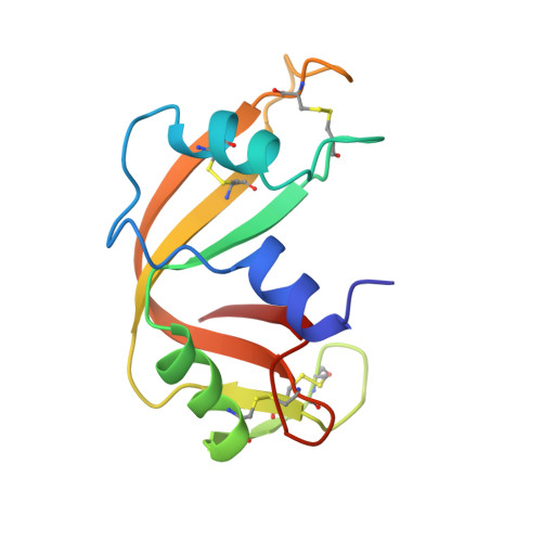
1QHC - PubMed Abstract:
The crystal structure of ribonuclease A (RNase A) in complex with pdUppA-3'-p [5'-phospho-2'-deoxyuridine-3'-pyrophosphate (P'-->5') adenosine 3'-phosphate] has been determined at 1.7 A resolution. This dinucleotide is the most potent low molecular weight inhibitor of RNase A reported to date (K(i) = 27 nM) and is also effective against two major nonpancreatic RNases: eosinophil-derived neurotoxin and RNase-4; in all cases, tight binding in large part derives from the unusual 3',5'-pyrophosphate internucleotide linkage [Russo, N., and Shapiro, R. (1999) J. Biol. Chem. 274, 14902-14908]. The design of pdUppA-3'-p was based on the crystal structure of RNase A complexed with 5'-diphosphoadenosine 3'-phosphate (ppA-3'-p) [Leonidas, D. D., Shapiro, R., Irons, L. I., Russo, N., and Acharya, K. R. (1997) Biochemistry 36, 5578-5588]. The adenosine of pdUppA-3'-p adopts an atypical syn conformation not observed for standard adenosine nucleotides bound to RNase A. This conformation, which allows extensive interactions with Asn 67, Gln 69, Asn 71, and His 119, is associated with the placement of the 5'-beta-phosphate of the adenylate, rather than alpha-phosphate, at the site where substrate phosphodiester bond cleavage occurs. The contacts of the deoxyuridine 5'-phosphate portion of pdUppA-3'-p appear to be responsible for the 9-fold increased affinity of this compound as compared to ppA-3'-p: the uracil base binds to Thr 45 in the same manner as previous pyrimidine inhibitors, and the terminal 5'-phosphate is positioned to form medium-range Coulombic interactions with Lys 66. The full potential benefit of these added interactions is not realized because of compensatory losses of hydrogen bonds of Lys 7 and Gln 11 with the terminal 3'-phosphate and the adenylate 5'-alpha-phosphate, which were not predicted by modeling. The results reported here have important implications for the design of improved inhibitors of RNase A and for the development of therapeutic agents to control the activities of RNase homologues such as eosinophil-derived neurotoxin and angiogenin that have roles in human pathologies.
Organizational Affiliation:
Department of Biology and Biochemistry, University of Bath, UK.