1QGJ

ARABIDOPSIS THALIANA PEROXIDASE N


Experimental Data Snapshot

  • Method: X-RAY DIFFRACTION
  • Resolution: 1.90 Å
  • R-Value Free: 0.205 
  • R-Value Work: 0.170 
  • R-Value Observed: 0.170 

wwPDB Validation   3D Report Full Report


Ligand Structure Quality Assessment 


This is version 1.3 of the entry. See complete history


Literature

Arabidopsis thaliana peroxidase N: structure of a novel neutral peroxidase.

Mirza, O.Henriksen, A.Ostergaard, L.Welinder, K.G.Gajhede, M.

(2000) Acta Crystallogr D Biol Crystallogr 56: 372-375

  • DOI: https://doi.org/10.1107/s0907444999016340
  • Primary Citation of Related Structures:  
    1QGJ

  • PubMed Abstract: 

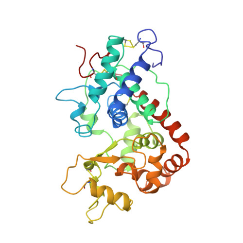
    The structure of the neutral peroxidase from Arabidopsis thaliana (ATP N) has been determined to a resolution of 1.9 A and a free R value of 20.5%. ATP N has the expected characteristic fold of the class III peroxidases, with a C(alpha) r.m.s.d. of 0.82 A when compared with horseradish peroxidase C (HRP C). HRP C is 54% identical to ATP N in sequence. When the structures of four class III plant peroxidases are superimposed, the regions with structural differences are non-randomly distributed; all are located in one half of the molecule. The architecture of the haem pocket of ATP N is very similar to that of HRP C, in agreement with the low small-molecule substrate specificity of all class III peroxidases. The structure of ATP N suggests that the pH dependence of the substrate turnover will differ from that of HRP C owing to differences in polarity of the residues in the substrate-access channel. Since there are fewer hydrogen bonds to haem C17 propionate O atoms in ATP N than in HRP C, it is suggested that ATP N will lose haem more easily than HRP C. Unlike almost all other class III plant peroxidases, ATP N has a free cysteine residue at a similar position to the suggested secondary substrate-binding site in lignin peroxidase.


  • Organizational Affiliation

    Protein Structure Group, Department of Chemistry, University of Copenhagen, Universitetsparken 5, DK-2100 Kobenhavn O, Denmark.


Macromolecules
Find similar proteins by:  (by identity cutoff)  |  3D Structure
Entity ID: 1
MoleculeChains Sequence LengthOrganismDetailsImage
PEROXIDASE N
A, B
300Arabidopsis thalianaMutation(s): 0 
EC: 1.11.1
UniProt
Find proteins for Q39034 (Arabidopsis thaliana)
Explore Q39034 
Go to UniProtKB:  Q39034
Entity Groups  
Sequence Clusters30% Identity50% Identity70% Identity90% Identity95% Identity100% Identity
UniProt GroupQ39034
Sequence Annotations
Expand
  • Reference Sequence
Small Molecules
Ligands 3 Unique
IDChains Name / Formula / InChI Key2D Diagram3D Interactions
HEM
Query on HEM

Download Ideal Coordinates CCD File 
E [auth A],
I [auth B]
PROTOPORPHYRIN IX CONTAINING FE
C34 H32 Fe N4 O4
KABFMIBPWCXCRK-RGGAHWMASA-L
GSH
Query on GSH

Download Ideal Coordinates CCD File 
F [auth A],
J [auth B]
GLUTATHIONE
C10 H17 N3 O6 S
RWSXRVCMGQZWBV-WDSKDSINSA-N
CA
Query on CA

Download Ideal Coordinates CCD File 
C [auth A],
D [auth A],
G [auth B],
H [auth B]
CALCIUM ION
Ca
BHPQYMZQTOCNFJ-UHFFFAOYSA-N
Experimental Data & Validation

Experimental Data

  • Method: X-RAY DIFFRACTION
  • Resolution: 1.90 Å
  • R-Value Free: 0.205 
  • R-Value Work: 0.170 
  • R-Value Observed: 0.170 
  • Space Group: P 21 21 21
Unit Cell:
Length ( Å )Angle ( ˚ )
a = 72.989α = 90
b = 87.457β = 90
c = 91.371γ = 90
Software Package:
Software NamePurpose
CNSrefinement

Structure Validation

View Full Validation Report


Ligand Structure Quality Assessment 


Entry History 

Deposition Data

Revision History  (Full details and data files)

  • Version 1.0: 2000-03-08
    Type: Initial release
  • Version 1.1: 2008-04-26
    Changes: Version format compliance
  • Version 1.2: 2011-07-13
    Changes: Version format compliance
  • Version 1.3: 2023-12-27
    Changes: Data collection, Database references, Derived calculations