Crystal structure of an inhibitor complex of the 3C proteinase from hepatitis A virus (HAV) and implications for the polyprotein processing in HAV.
Bergmann, E.M., Cherney, M.M., Mckendrick, J., Frormann, S., Luo, C., Malcolm, B.A., Vederas, J.C., James, M.N.(1999) Virology 265: 153-163
- PubMed: 10603326
- DOI: https://doi.org/10.1006/viro.1999.9968
- Primary Citation of Related Structures:
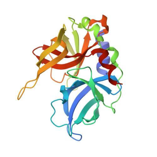
1QA7 - PubMed Abstract:
The proteolytic processing of the viral polyprotein is an essential step during the life cycle of hepatitis A virus (HAV), as it is in all positive-sense, single-stranded RNA viruses of animals. In HAV the 3C proteinase is the only proteolytic activity involved in the polyprotein processing. The specific recognition of the cleavage sites by the 3C proteinase depends on the amino acid sequence of the cleavage site. The structure of the complex of the HAV 3C proteinase and a dipeptide inhibitor has been determined by X-ray crystallography. The double-mutant of HAV 3C (C24S, F82A) was inhibited with the specific inhibitor iodoacetyl-valyl-phenylalanyl-amide. The resulting complex had an acetyl-Val-Phe-amide group covalently attached to the S(gamma) atom of the nucleophilic Cys 172 of the enzyme. Crystals of the complex of HAV 3C (C24S, F82A) acetyl-Val-Phe-amide were found to be monoclinic, space group P2(1), having 4 molecules in the asymmetric unit and diffracting to 1.9-A resolution. The final refined structure consists of 4 molecules of HAV 3C (C24S,F82A) acetyl-Val-Phe-amide, 1 molecule of DMSO, 1 molecule of glycerol, and 514 water molecules. There are considerable conformational differences among the four molecules in the asymmetric unit. The final R-factor is 20.4% for all observed reflections between 15.0- and 1.9-A resolution and the corresponding R(free) is 29.8%. The dipeptide inhibitor is bound to the S(1)(') and S(2)(') specificity subsites of the proteinase. The crystal structure reveals that the HAV 3C proteinase possesses a well-defined S(2)(') specificity pocket and suggests that the P(2)(') residue could be an important determinant for the selection of the primary cleavage site during the polyprotein processing in HAV.
Organizational Affiliation:
Department of Biochemistry, University of Alberta, Edmonton, Alberta, T6G 2H7, Canada.