1Q9H

3-Dimensional structure of native Cel7A from Talaromyces emersonii


Experimental Data Snapshot

  • Method: X-RAY DIFFRACTION
  • Resolution: 2.35 Å
  • R-Value Free: 0.229 
  • R-Value Work: 0.161 
  • R-Value Observed: 0.165 

wwPDB Validation   3D Report Full Report


Ligand Structure Quality Assessment 


This is version 3.0 of the entry. See complete history


Literature

Three-dimensional structure of a thermostable native cellobiohydrolase, CBH IB, and molecular characterization of the cel7 gene from the filamentous fungus, Talaromyces emersonii

Grassick, A.Murray, P.G.Thompson, R.Collins, C.M.Byrnes, L.Birrane, G.Higgins, T.M.Tuohy, M.G.

(2004) Eur J Biochem 271: 4495-4506

  • DOI: https://doi.org/10.1111/j.1432-1033.2004.04409.x
  • Primary Citation of Related Structures:  
    1Q9H

  • PubMed Abstract: 

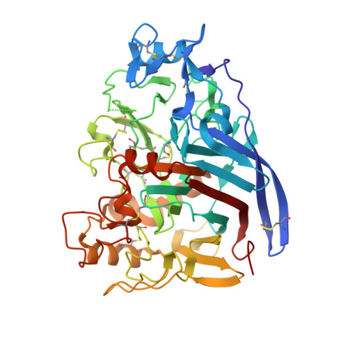
    The X-ray structure of native cellobiohydrolase IB (CBH IB) from the filamentous fungus Talaromyces emersonii, PDB 1Q9H, was solved to 2.4 A by molecular replacement. 1Q9H is a glycoprotein that consists of a large, single domain with dimensions of approximately 60 A x 40 A x 50 A and an overall beta-sandwich structure, the characteristic fold of Family 7 glycosyl hydrolases (GH7). It is the first structure of a native glycoprotein and cellulase from this thermophilic eukaryote. The long cellulose-binding tunnel seen in GH7 Cel7A from Trichoderma reesei is conserved in 1Q9H, as are the catalytic residues. As a result of deletions and other changes in loop regions, the binding and catalytic properties of T. emersonii 1Q9H are different. The gene (cel7) encoding CBH IB was isolated from T. emersonii and expressed heterologously with an N-terminal polyHis-tag, in Escherichia coli. The deduced amino acid sequence of cel7 is homologous to fungal cellobiohydrolases in GH7. The recombinant cellobiohydrolase was virtually inactive against methylumberiferyl-cellobioside and chloronitrophenyl-lactoside, but partial activity could be restored after refolding of the urea-denatured enzyme. Profiles of cel7 expression in T. emersonii, investigated by Northern blot analysis, revealed that expression is regulated at the transcriptional level. Putative regulatory element consensus sequences for cellulase transcription factors have been identified in the upstream region of the cel7 genomic sequence.


  • Organizational Affiliation

    Molecular Glycobiotechnology Group, Department of Biochemistry, National University of Ireland, Galway, Ireland.


Macromolecules
Find similar proteins by:  (by identity cutoff)  |  3D Structure
Entity ID: 1
MoleculeChains Sequence LengthOrganismDetailsImage
cellobiohydrolase I catalytic domain437Rasamsonia emersoniiMutation(s): 0 
EC: 3.2.1.91
UniProt
Find proteins for Q8TFL9 (Talaromyces emersonii)
Explore Q8TFL9 
Go to UniProtKB:  Q8TFL9
Entity Groups  
Sequence Clusters30% Identity50% Identity70% Identity90% Identity95% Identity100% Identity
UniProt GroupQ8TFL9
Sequence Annotations
Expand
  • Reference Sequence
Oligosaccharides

Help

Entity ID: 2
MoleculeChains Length2D Diagram Glycosylation3D Interactions
2-acetamido-2-deoxy-beta-D-glucopyranose-(1-4)-2-acetamido-2-deoxy-beta-D-glucopyranose
B
2N-Glycosylation
Glycosylation Resources
GlyTouCan:  G42666HT
GlyCosmos:  G42666HT
GlyGen:  G42666HT
Small Molecules
Ligands 1 Unique
IDChains Name / Formula / InChI Key2D Diagram3D Interactions
NAG
Query on NAG

Download Ideal Coordinates CCD File 
C [auth A]2-acetamido-2-deoxy-beta-D-glucopyranose
C8 H15 N O6
OVRNDRQMDRJTHS-FMDGEEDCSA-N
Modified Residues  1 Unique
IDChains TypeFormula2D DiagramParent
PCA
Query on PCA
A
L-PEPTIDE LINKINGC5 H7 N O3GLN
Experimental Data & Validation

Experimental Data

  • Method: X-RAY DIFFRACTION
  • Resolution: 2.35 Å
  • R-Value Free: 0.229 
  • R-Value Work: 0.161 
  • R-Value Observed: 0.165 
  • Space Group: P 41 21 2
Unit Cell:
Length ( Å )Angle ( ˚ )
a = 74.395α = 90
b = 74.395β = 90
c = 177.046γ = 90
Software Package:
Software NamePurpose
REFMACrefinement
DENZOdata reduction
SCALEPACKdata scaling
AMoREphasing

Structure Validation

View Full Validation Report


Ligand Structure Quality Assessment 


Entry History 

Deposition Data

Revision History  (Full details and data files)

  • Version 1.0: 2004-11-09
    Type: Initial release
  • Version 1.1: 2008-04-29
    Changes: Version format compliance
  • Version 1.2: 2011-07-13
    Changes: Non-polymer description, Version format compliance
  • Version 2.0: 2019-12-25
    Changes: Data collection, Database references, Derived calculations, Polymer sequence
  • Version 3.0: 2020-07-29
    Type: Remediation
    Reason: Carbohydrate remediation
    Changes: Atomic model, Data collection, Derived calculations, Structure summary