1Q8F

Crystal Structure of the E.coli pyrimidine nucleoside hydrolase yeiK


Experimental Data Snapshot

  • Method: X-RAY DIFFRACTION
  • Resolution: 1.70 Å
  • R-Value Free: 0.170 
  • R-Value Work: 0.153 
  • R-Value Observed: 0.153 

wwPDB Validation   3D Report Full Report


This is version 1.3 of the entry. See complete history


Literature

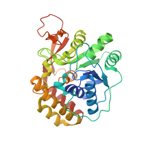
Crystal structure to 1.7 a of the Escherichia coli pyrimidine nucleoside hydrolase YeiK, a novel candidate for cancer gene therapy.

Giabbai, B.Degano, M.

(2004) Structure 12: 739-749

  • DOI: https://doi.org/10.1016/j.str.2004.03.018
  • Primary Citation of Related Structures:  
    1Q8F

  • PubMed Abstract: 

    Enzymes with nucleoside hydrolase (NH) activity are crucial for salvaging nucleic acid components in purine auxotrophic protozoan parasites, but are also present in prokaryotes and higher eukaryotes. Here we analyze the distribution of genes encoding for putative NH proteins and characterize the yeiK gene product from Escherichia coli as a pyrimidine-specific NH. The crystal structure of YeiK to 1.7 A defines the structural basis for its substrate specificity and identifies residues involved in the catalytic mechanism that differ from both nonspecific and purine-specific NHs. Large differences in the tetrameric quaternary structure compared to nonspecific protozoan NHs are brought forth by minor differences in the interacting surfaces. The first structural and functional characterization of a nonparasitic, pyrimidine nucleoside-specific NH suggests a possible role for these enzymes in the metabolism of tRNA nucleosides. The high catalytic efficiency of YeiK toward 5-fluorouridine could be exploited for suicide gene therapy in cancer treatment.


  • Organizational Affiliation

    Biocrystallography Unit, DIBIT, San Raffaele Scientific Institute, via Olgettina 58, I-20132 Milan, Italy.


Macromolecules
Find similar proteins by:  (by identity cutoff)  |  3D Structure
Entity ID: 1
MoleculeChains Sequence LengthOrganismDetailsImage
Pyrimidine nucleoside hydrolase
A, B, C, D
313Escherichia coliMutation(s): 0 
Gene Names: YEIK
EC: 3.2.2.8
UniProt
Find proteins for P33022 (Escherichia coli (strain K12))
Explore P33022 
Go to UniProtKB:  P33022
Entity Groups  
Sequence Clusters30% Identity50% Identity70% Identity90% Identity95% Identity100% Identity
UniProt GroupP33022
Sequence Annotations
Expand
  • Reference Sequence
Small Molecules
Ligands 2 Unique
IDChains Name / Formula / InChI Key2D Diagram3D Interactions
GOL
Query on GOL

Download Ideal Coordinates CCD File 
F [auth A]
G [auth A]
H [auth A]
I [auth A]
J [auth A]
F [auth A],
G [auth A],
H [auth A],
I [auth A],
J [auth A],
L [auth B],
M [auth B],
N [auth B],
P [auth C],
Q [auth C],
R [auth C],
S [auth C],
U [auth D],
V [auth D],
W [auth D],
X [auth D]
GLYCEROL
C3 H8 O3
PEDCQBHIVMGVHV-UHFFFAOYSA-N
CA
Query on CA

Download Ideal Coordinates CCD File 
E [auth A],
K [auth B],
O [auth C],
T [auth D]
CALCIUM ION
Ca
BHPQYMZQTOCNFJ-UHFFFAOYSA-N
Experimental Data & Validation

Experimental Data

  • Method: X-RAY DIFFRACTION
  • Resolution: 1.70 Å
  • R-Value Free: 0.170 
  • R-Value Work: 0.153 
  • R-Value Observed: 0.153 
  • Space Group: P 1
Unit Cell:
Length ( Å )Angle ( ˚ )
a = 44.808α = 112.95
b = 85.707β = 101.95
c = 90.678γ = 85.92
Software Package:
Software NamePurpose
REFMACrefinement
MOSFLMdata reduction
CCP4data scaling
MOLREPphasing

Structure Validation

View Full Validation Report


Entry History 

Deposition Data

Revision History  (Full details and data files)

  • Version 1.0: 2004-05-11
    Type: Initial release
  • Version 1.1: 2008-04-29
    Changes: Version format compliance
  • Version 1.2: 2011-07-13
    Changes: Advisory, Version format compliance
  • Version 1.3: 2023-08-16
    Changes: Data collection, Database references, Derived calculations, Refinement description