Crystal structure of a C-terminal deletion mutant of human protein kinase CK2 catalytic subunit
Ermakova, I., Boldyreff, B., Issinger, O.-G., Niefind, K.(2003) J Mol Biol 330: 925-934
- PubMed: 12860116
- DOI: https://doi.org/10.1016/s0022-2836(03)00638-7
- Primary Citation of Related Structures:
1PJK - PubMed Abstract:
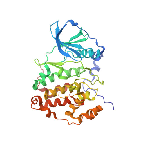
Protein kinase CK2 (formerly called: casein kinase 2) is a heterotetrameric enzyme composed of two separate catalytic chains (CK2alpha) and a stable dimer of two non-catalytic subunits (CK2beta). CK2alpha is a highly conserved member of the superfamily of eukaryotic protein kinases. The crystal structure of a C-terminal deletion mutant of human CK2alpha was solved and refined to 2.5A resolution. In the crystal the CK2alpha mutant exists as a monomer in agreement with the organization of the subunits in the CK2 holoenzyme. The refined structure shows the helix alphaC and the activation segment, two main regions of conformational plasticity and regulatory importance in eukaryotic protein kinases, in active conformations stabilized by extensive contacts to the N-terminal segment. This arrangement is in accordance with the constitutive activity of the enzyme. By structural superimposition of human CK2alpha in isolated form and embedded in the human CK2 holoenzyme the loop connecting the strands beta4 and beta5 and the ATP-binding loop were identified as elements of structural variability. This structural comparison suggests that the ATP-binding loop may be the key region by which the non-catalytic CK2beta dimer modulates the activity of CK2alpha. The beta4/beta5 loop was found in a closed conformation in contrast to the open conformation observed for the CK2alpha subunits of the CK2 holoenzyme. CK2alpha monomers with this closed beta4/beta5 loop conformation are unable to bind CK2beta dimers in the common way for sterical reasons, suggesting a mechanism to protect CK2alpha from integration into CK2 holoenzyme complexes. This observation is consistent with the growing evidence that CK2alpha monomers and CK2beta dimers can exist in vivo independently from the CK2 holoenzyme and may possess physiological roles of their own.
Organizational Affiliation:
Universität zu Köln, Institut für Biochemie, Zülpicher Strasse 47, D-50674 Köln, Germany.