1PBY

Structure of the Phenylhydrazine Adduct of the Quinohemoprotein Amine Dehydrogenase from Paracoccus denitrificans at 1.7 A Resolution


Experimental Data Snapshot

  • Method: X-RAY DIFFRACTION
  • Resolution: 1.70 Å
  • R-Value Free: 0.224 
  • R-Value Work: 0.191 

wwPDB Validation   3D Report Full Report


Ligand Structure Quality Assessment 


This is version 2.1 of the entry. See complete history


Literature

Structure of the phenylhydrazine adduct of the quinohemoprotein amine dehydrogenase from Paracoccus denitrificans at 1.7 A resolution.

Datta, S.Ikeda, T.Kano, K.Mathews, F.S.

(2003) Acta Crystallogr D Biol Crystallogr 59: 1551-1556

  • DOI: https://doi.org/10.1107/s090744490301429x
  • Primary Citation of Related Structures:  
    1PBY

  • PubMed Abstract: 

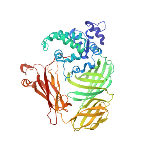
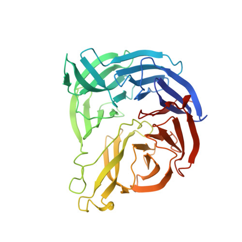
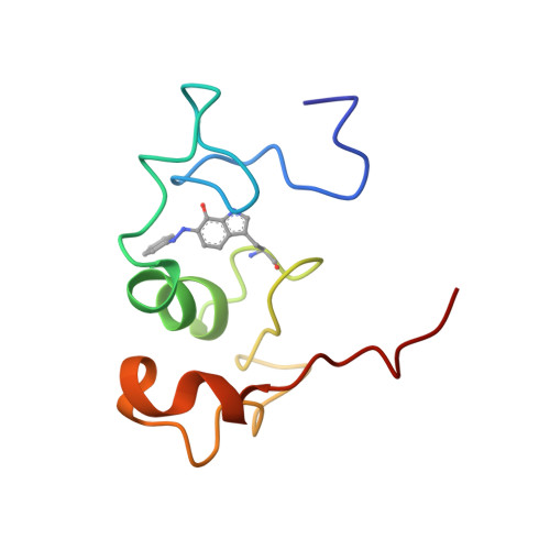
    The 109 kDa quinohemoprotein amine dehydrogenase (QHNDH) from Paracoccus denitrificans contains a novel redox cofactor, cysteine tryptophylquinone (CTQ). This cofactor is derived from a pair of gene-encoded amino acids by post-translational modification and was previously identified and characterized within an 82-residue subunit by chemical methods and crystallographic analysis at 2.05 A resolution. It contains an orthoquinone moiety bound to the indole ring and catalyzes the oxidation of aliphatic and aromatic amines through formation of a Schiff-base intermediate involving one of the quinone O atoms. This paper reports the structural analysis of the complex of QHNDH with the enzyme inhibitor phenylhydrazine determined at 1.70 A resolution. The phenylhydrazone product is attached to the C6 position, identifying the O6 atom of CTQ as the site of Schiff-base formation as postulated by analogy to another amine-oxidizing enzyme, methylamine dehydrogenase. Furthermore, the inner N atom closest to the phenyl ring of phenylhydrazine forms a hydrogen bond to gammaAsp33 in the complex, lending support to the hypothesis that this residue serves as the active-site base for proton abstraction during catalysis.


  • Organizational Affiliation

    Washington University School of Medicine, St Louis, MO 63110, USA.


Macromolecules
Find similar proteins by:  (by identity cutoff)  |  3D Structure
Entity ID: 1
MoleculeChains Sequence LengthOrganismDetailsImage
quinohemoprotein amine dehydrogenase 60 kDa subunit489Paracoccus denitrificansMutation(s): 0 
EC: 1.4.99.3
UniProt
Find proteins for Q8VUT0 (Paracoccus denitrificans)
Explore Q8VUT0 
Go to UniProtKB:  Q8VUT0
Entity Groups  
Sequence Clusters30% Identity50% Identity70% Identity90% Identity95% Identity100% Identity
UniProt GroupQ8VUT0
Sequence Annotations
Expand
  • Reference Sequence
Find similar proteins by:  (by identity cutoff)  |  3D Structure
Entity ID: 2
MoleculeChains Sequence LengthOrganismDetailsImage
quinohemoprotein amine dehydrogenase 40 kDa subunit337Paracoccus denitrificansMutation(s): 0 
UniProt
Find proteins for Q8VUS7 (Paracoccus denitrificans)
Explore Q8VUS7 
Go to UniProtKB:  Q8VUS7
Entity Groups  
Sequence Clusters30% Identity50% Identity70% Identity90% Identity95% Identity100% Identity
UniProt GroupQ8VUS7
Sequence Annotations
Expand
  • Reference Sequence
Find similar proteins by:  (by identity cutoff)  |  3D Structure
Entity ID: 3
MoleculeChains Sequence LengthOrganismDetailsImage
quinohemoprotein amine dehydrogenase 9 kDa subunit79Paracoccus denitrificansMutation(s): 1 
UniProt
Find proteins for Q8VUS8 (Paracoccus denitrificans)
Explore Q8VUS8 
Go to UniProtKB:  Q8VUS8
Entity Groups  
Sequence Clusters30% Identity50% Identity70% Identity90% Identity95% Identity100% Identity
UniProt GroupQ8VUS8
Sequence Annotations
Expand
  • Reference Sequence
Experimental Data & Validation

Experimental Data

  • Method: X-RAY DIFFRACTION
  • Resolution: 1.70 Å
  • R-Value Free: 0.224 
  • R-Value Work: 0.191 
  • Space Group: P 41 21 2
Unit Cell:
Length ( Å )Angle ( ˚ )
a = 99.237α = 90
b = 99.237β = 90
c = 213.056γ = 90
Software Package:
Software NamePurpose
CNSrefinement
DENZOdata reduction
SCALEPACKdata scaling

Structure Validation

View Full Validation Report


Ligand Structure Quality Assessment 


Entry History 

Deposition Data

Revision History  (Full details and data files)

  • Version 1.0: 2003-09-02
    Type: Initial release
  • Version 1.1: 2008-04-29
    Changes: Version format compliance
  • Version 1.2: 2011-07-13
    Changes: Version format compliance
  • Version 1.3: 2017-10-11
    Changes: Refinement description
  • Version 2.0: 2021-03-03
    Changes: Advisory, Atomic model, Data collection, Database references, Derived calculations, Non-polymer description, Structure summary
  • Version 2.1: 2023-08-16
    Changes: Data collection, Database references, Refinement description