Crystal structure of the p-hydroxybenzoate hydroxylase-substrate complex refined at 1.9 A resolution. Analysis of the enzyme-substrate and enzyme-product complexes.
Schreuder, H.A., Prick, P.A., Wierenga, R.K., Vriend, G., Wilson, K.S., Hol, W.G., Drenth, J.(1989) J Mol Biol 208: 679-696
- PubMed: 2553983
- DOI: https://doi.org/10.1016/0022-2836(89)90158-7
- Primary Citation of Related Structures:
1PBE - PubMed Abstract:
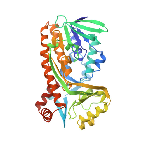
Using synchrotron radiation, the X-ray diffraction intensities of crystals of p-hydroxy-benzoate hydroxylase, complexed with the substrate p-hydroxybenzoate, were measured to a resolution of 1.9 A. Restrained least-squares refinement alternated with rebuilding in electron density maps yielded an atom model of the enzyme-substrate complex with a crystallographic R-factor of 15.6% for 31,148 reflections between 6.0 and 1.9 A. A total of 330 solvent molecules was located. In the final model, only three residues have deviating phi-psi angle combinations. One of them, the active site residue Arg44, has a well-defined electron density and may be strained to adopt this conformation for efficient catalysis. The mode of binding of FAD is distinctly different for the different components of the coenzyme. The adenine ring is engaged in three water-mediated hydrogen bonds with the protein, while making only one direct hydrogen bond with the enzyme. The pyrophosphate moiety makes five water-mediated versus three direct hydrogen bonds. The ribityl and ribose moieties make only direct hydrogen bonds, in all cases, except one, with side-chain atoms. The isoalloxazine ring also makes only direct hydrogen bonds, but virtually only with main-chain atoms. The conformation of FAD in p-hydroxybenzoate hydroxylase is strikingly similar to that in glutathione reductase, while the riboflavin-binding parts of these two enzymes have no structural similarity at all. The refined 1.9 A structure of the p-hydroxybenzoate hydroxylase-substrate complex was the basis of further refinement of the 2.3 A structure of the enzyme-product complex. The result was a final R-factor of 16.7% for 14,339 reflections between 6.0 and 2.3 A and an improved geometry. Comparison between the complexes indicated only small differences in the active site region, where the product molecule is rotated by 14 degrees compared with the substrate in the enzyme-substrate complex. During the refinements of the enzyme-substrate and enzyme-product complexes, the flavin ring was allowed to bend or twist by imposing planarity restraints on the benzene and pyrimidine ring, but not on the flavin ring as a whole. The observed angle between the benzene ring and the pyrimidine ring was 10 degrees for the enzyme-substrate complex and 19 degrees for the enzyme-product complex. Because of the high temperature factors of the flavin ring in the enzyme-product complex, the latter value should be treated with caution. Six out of eight peptide residues near the flavin ring are oriented with their nitrogen atom pointing towards the ring.(ABSTRACT TRUNCATED AT 400 WORDS)
Organizational Affiliation:
Laboratory of Chemical Physics, University of Groningen, The Netherlands.