NMR structure determination of a novel conotoxin, [Pro 7,13] alpha A-conotoxin PIVA.
Han, K.H., Hwang, K.J., Kim, S.M., Kim, S.K., Gray, W.R., Olivera, B.M., Rivier, J., Shon, K.J.(1997) Biochemistry 36: 1669-1677
- PubMed: 9048550
- DOI: https://doi.org/10.1021/bi962301k
- Primary Citation of Related Structures:
1P1P - PubMed Abstract:
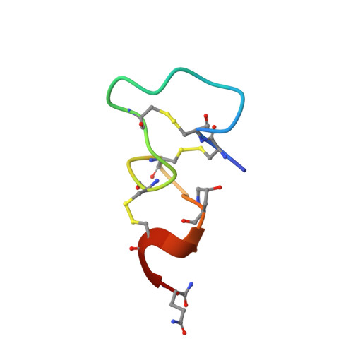
A high-resolution solution conformation of a novel conotoxin, [Pro 7,13] alpha A-conotoxin PIVA, GCCGSYPNAACHPCSCKDROSYCGQ-NH2, has been determined by two-dimensional 1H NMR methods and distance geometry calculations. The total of 324 NOE-derived interproton distance restraints including 33 long-range NOE restraints as well as 11 phi and 7 chi 1 torsion angle restraints was used for computation of structures. Back-calculation from the experimental NOE spectrum has provided 49 new NOE restraints and yielded the final R-factors of Ra = 0.641 and Rb = 0.157. The final RMSD values are 0.90 and 1.16 A for the backbone and the heavy atoms, respectively. The C-terminal half of the molecule involving the residues 12-24 is extremely well-defined with a backbone RMSD value of 0.56 A, whereas the N-terminal 3-11 disulfide loop is relatively flexible, possessing a backbone RMSD value of 1.09 A. The [Pro 7,13] alpha A-conotoxin PIVA does not contain any significant secondary structure although the 21S-24G nearly completes one turn of a 3(10) helix. The overall protein fold is largely maintained by the three disulfide bridges of 2-16, 3-11, and 14-23. The presence of the three disulfide bridges imposes geometric constraints that force the molecule to form six continuous bends involving the following residues: 3C-5S, 7P-10A, 12H-14C, 15S-17K, 17K-19R, and 21S-25Q. The overall shape of the [Pro 7,13] alpha A-conotoxin PIVA can be described as an "iron". Residues 15S-19R form a loop that protrudes out of the "bottom plate" formed by the rest of the protein and constitute the handle of the iron. The N-terminal tip of the molecule is relatively immobile due to attractive electrostatic interactions between the gamma-hydroxyl group of 20 Hyp and the phenolic hydroxyl group of 22Y. The flexible 3-11 disulfide loop consists mostly of hydrophobic residues, while the best-defined 14-23 disulfide loop contains the highly charged hydrophilic 15S-19R "handle" domain exposed to the exterior of the protein. Binding to nicotinic acetylcholine receptor can be mediated through two different types of interactions: one involving the aromatic hydrophobic residues such as 6Y and 12H and the other involving the positively charged hydrophilic side chain of the 19R. The side chain of the 19R in the [Pro 7, 13] alpha A-conotoxin PIVA and that of the 9R of the alpha-conotoxin G1, and also the side chains of the 12H and 6Y in the former and those of 10H and 11Y in the latter can be aligned to point to the same direction when the corresponding backbone atoms are superimposed to an RMSD value of 2.5 A.
Organizational Affiliation:
Biomolecular Structure Research Unit, Korea Research Institute of Bioscience and Biotechnology, Taejon, Korea. khhan@biotech5.geri.re.kr