1P1J

Crystal structure of the 1L-myo-inositol 1-phosphate synthase complexed with NADH


Experimental Data Snapshot

  • Method: X-RAY DIFFRACTION
  • Resolution: 1.70 Å
  • R-Value Free: 0.190 
  • R-Value Work: 0.165 
  • R-Value Observed: 0.167 

wwPDB Validation   3D Report Full Report


Ligand Structure Quality Assessment 


This is version 1.3 of the entry. See complete history


Literature

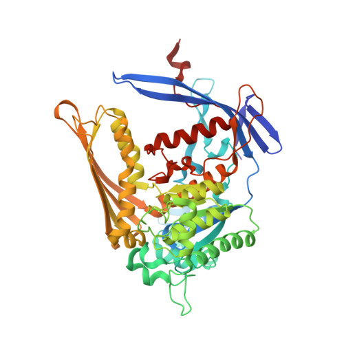
Structures of NAD(+)- and NADH-bound 1-l-myo-inositol 1-phosphate synthase.

Jin, X.Geiger, J.H.

(2003) Acta Crystallogr D Biol Crystallogr 59: 1154-1164

  • DOI: https://doi.org/10.1107/s0907444903008205
  • Primary Citation of Related Structures:  
    1P1F, 1P1H, 1P1I, 1P1J, 1P1K

  • PubMed Abstract: 

    1-l-myo-Inositol 1-phosphate synthase catalyzes the conversion of d-glucose 6-phosphate to 1-l-myo-inositol 1-phosphate, the first and rate-limiting step in the biosynthesis of all inositol-containing compounds. It involves an oxidation, an intramolecular aldol cyclization and a reduction. Here, the structure of the enzyme in its NAD(+)-bound, NADH-bound and apo forms is presented. These structures confirm that a significant portion of the active site is disordered in the absence of a small molecule, as none of the NAD(+)-bound forms of the enzyme have ordered active sites. On the other hand, the NADH-bound form contains two small molecules in the active site: a phosphate and glycerol. The entire active site is ordered in the presence of these two molecules, completely encapsulating them within the interior cavity. Significant changes in the structure of the active site are also seen, including repositioning of the nicotinamide ring and a motion of a loop region to accommodate the bound phosphate. These changes call into question the mechanism previously proposed for the enzyme. A comparison of the yeast and mycobacterial enzymes shows a surprisingly large change in the relative orientation of the catalytic and Rossmann-fold domains in the two enzymes.


  • Organizational Affiliation

    Department of Chemistry, Michigan State University, USA.


Macromolecules
Find similar proteins by:  (by identity cutoff)  |  3D Structure
Entity ID: 1
MoleculeChains Sequence LengthOrganismDetailsImage
Inositol-3-phosphate synthase
A, B
533Saccharomyces cerevisiaeMutation(s): 0 
EC: 5.5.1.4
UniProt
Find proteins for P11986 (Saccharomyces cerevisiae (strain ATCC 204508 / S288c))
Explore P11986 
Go to UniProtKB:  P11986
Entity Groups  
Sequence Clusters30% Identity50% Identity70% Identity90% Identity95% Identity100% Identity
UniProt GroupP11986
Sequence Annotations
Expand
  • Reference Sequence
Experimental Data & Validation

Experimental Data

  • Method: X-RAY DIFFRACTION
  • Resolution: 1.70 Å
  • R-Value Free: 0.190 
  • R-Value Work: 0.165 
  • R-Value Observed: 0.167 
  • Space Group: C 1 2 1
Unit Cell:
Length ( Å )Angle ( ˚ )
a = 149.86α = 90
b = 99.851β = 126.64
c = 122.571γ = 90
Software Package:
Software NamePurpose
HKL-2000data collection
SCALEPACKdata scaling
CNSrefinement
HKL-2000data reduction
CNSphasing

Structure Validation

View Full Validation Report


Ligand Structure Quality Assessment 


Entry History 

Deposition Data

Revision History  (Full details and data files)

  • Version 1.0: 2003-07-08
    Type: Initial release
  • Version 1.1: 2008-04-29
    Changes: Version format compliance
  • Version 1.2: 2011-07-13
    Changes: Derived calculations, Version format compliance
  • Version 1.3: 2024-02-14
    Changes: Data collection, Database references, Derived calculations