1OQL

Mistletoe Lectin I from Viscum album complexed with galactose


Experimental Data Snapshot

  • Method: X-RAY DIFFRACTION
  • Resolution: 3.00 Å
  • R-Value Free: 0.240 
  • R-Value Work: 0.199 
  • R-Value Observed: 0.201 

wwPDB Validation   3D Report Full Report


Ligand Structure Quality Assessment 


This is version 2.1 of the entry. See complete history


Literature

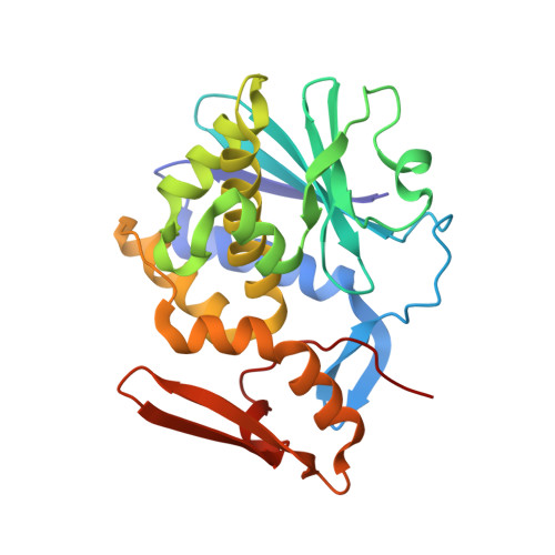
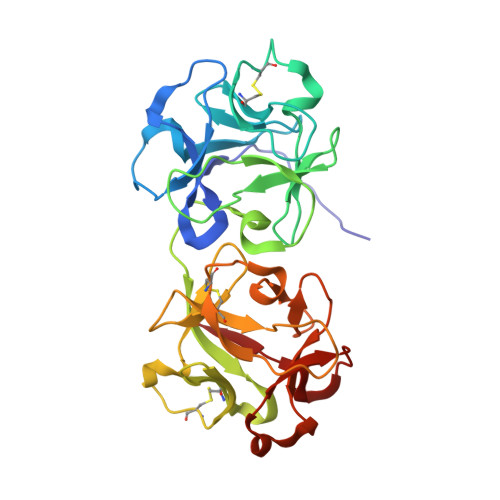
Crystal structure at 3 A of mistletoe lectin I, a dimeric type-II ribosome-inactivating protein, complexed with galactose

Niwa, H.Tonevitsky, A.G.Agapov, I.I.Saward, S.Pfuller, U.Palmer, R.A.

(2003) Eur J Biochem 270: 2739-2749

  • DOI: https://doi.org/10.1046/j.1432-1033.2003.03646.x
  • Primary Citation of Related Structures:  
    1OQL

  • PubMed Abstract: 

    The X-ray structure of mistletoe lectin I (MLI), a type-II ribosome-inactivating protein (RIP), cocrystallized with galactose is described. The model was refined at 3.0 A resolution to an R-factor of 19.9% using 21 899 reflections, with Rfree 24.0%. MLI forms a homodimer (A-B)2 in the crystal, as it does in solution at high concentration. The dimer is formed through contacts between the N-terminal domains of two B-chains involving weak polar and non-polar interactions. Consequently, the overall arrangement of sugar-binding sites in MLI differs from those in monomeric type-II RIPs: two N-terminal sugar-binding sites are 15 A apart on one side of the dimer, and two C-terminal sugar-binding sites are 87 A apart on the other side. Galactose binding is achieved by common hydrogen bonds for the two binding sites via hydroxy groups 3-OH and 4-OH and hydrophobic contact by an aromatic ring. In addition, at the N-terminal site 2-OH forms hydrogen bonds with Asp27 and Lys41, and at the C-terminal site 3-OH and 6-OH undergo water-mediated interactions and C5 has a hydrophobic contact. MLI is a galactose-specific lectin and shows little affinity for N-acetylgalactosamine. The reason for this is discussed. Structural differences among the RIPs investigated in this study (their quaternary structures, location of sugar-binding sites, and fine sugar specificities of their B-chains, which could have diverged through evolution from a two-domain protein) may affect the binding sites, and consequently the cellular transport processes and biological responses of these toxins.


  • Organizational Affiliation

    School of Crystallography, Birkbeck College, University of London, Malet Street, London WC1E 7HX, UK. h.niwa@mail.cryst.bbk.ac.uk


Macromolecules
Find similar proteins by:  (by identity cutoff)  |  3D Structure
Entity ID: 1
MoleculeChains Sequence LengthOrganismDetailsImage
MISTLETOE LECTIN I249Viscum albumMutation(s): 0 
EC: 3.2.2.22
UniProt
Find proteins for P81446 (Viscum album)
Explore P81446 
Go to UniProtKB:  P81446
Entity Groups  
Sequence Clusters30% Identity50% Identity70% Identity90% Identity95% Identity100% Identity
UniProt GroupP81446
Sequence Annotations
Expand
  • Reference Sequence
Find similar proteins by:  (by identity cutoff)  |  3D Structure
Entity ID: 2
MoleculeChains Sequence LengthOrganismDetailsImage
MISTLETOE LECTIN I263Viscum albumMutation(s): 0 
UniProt
Find proteins for P81446 (Viscum album)
Explore P81446 
Go to UniProtKB:  P81446
Entity Groups  
Sequence Clusters30% Identity50% Identity70% Identity90% Identity95% Identity100% Identity
UniProt GroupP81446
Sequence Annotations
Expand
  • Reference Sequence
Oligosaccharides

Help

Entity ID: 3
MoleculeChains Length2D Diagram Glycosylation3D Interactions
2-acetamido-2-deoxy-beta-D-glucopyranose-(1-4)-2-acetamido-2-deoxy-beta-D-glucopyranose
C, D
2N-Glycosylation
Glycosylation Resources
GlyTouCan:  G42666HT
GlyCosmos:  G42666HT
GlyGen:  G42666HT
Experimental Data & Validation

Experimental Data

  • Method: X-RAY DIFFRACTION
  • Resolution: 3.00 Å
  • R-Value Free: 0.240 
  • R-Value Work: 0.199 
  • R-Value Observed: 0.201 
  • Space Group: P 65 2 2
Unit Cell:
Length ( Å )Angle ( ˚ )
a = 107.65α = 90
b = 107.65β = 90
c = 311.92γ = 120
Software Package:
Software NamePurpose
CNSrefinement
DENZOdata reduction
CCP4data scaling
AMoREphasing

Structure Validation

View Full Validation Report


Ligand Structure Quality Assessment 


Entry History 

Deposition Data

Revision History  (Full details and data files)

  • Version 1.0: 2003-07-01
    Type: Initial release
  • Version 1.1: 2008-04-29
    Changes: Version format compliance
  • Version 1.2: 2011-07-13
    Changes: Derived calculations, Version format compliance
  • Version 2.0: 2020-07-29
    Type: Remediation
    Reason: Carbohydrate remediation
    Changes: Atomic model, Data collection, Derived calculations, Structure summary
  • Version 2.1: 2023-10-25
    Changes: Data collection, Database references, Refinement description, Structure summary