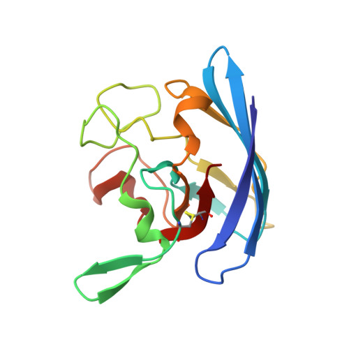
Active-Site Copper and Zinc Ions Modulate the Quaternary Structure of Prokaryotic Cu,Zn Superoxide Dismutase
Cioni, P., Pesce, A., Morozzo Della Rocca, B., Castelli, S., Falconi, M., Parrilli, L., Bolognesi, M., Strambini, G., Desideri, A.(2003) J Mol Biol 326: 1351
- PubMed: 12595249
- DOI: https://doi.org/10.1016/s0022-2836(03)00047-0
- Primary Citation of Related Structures:
1OAJ, 1OAL - PubMed Abstract:
The influence of the constitutive metal ions on the equilibrium properties of dimeric Photobacterium leiognathi Cu,Zn superoxide dismutase has been studied for the wild-type and for two mutant protein forms bearing a negative charge in the amino acid clusters at the dimer association interface. Depletion of copper and zinc dissociates the two mutant proteins into monomers, which reassemble toward the dimeric state upon addition of stoichiometric amounts of zinc. Pressure-dependent dissociation is observed for the copper-depleted wild-type and mutated enzymes, as monitored by the fluorescence shift of a unique tryptophan residue located at the subunit association interface. The spectral shift occurs slowly, reaching a plateau after 15-20 minutes, and is fully reversible. The recovery of the original fluorescence properties, after decompression, is fast (less than four minutes), suggesting that the isolated subunit has a relatively stable structure, and excluding the presence of stable intermediates during the dimer-monomer transition. The dimer dissociation process is still incomplete at 6.5 kbar for the copper-depleted wild-type and mutated enzymes, at variance with what is generally observed for oligomeric proteins that dissociate below 3 kbar. Measurement of the degree of dissociation, at two different protein concentrations, allows us to calculate the standard volume variation upon association, Delta V, and the dissociation constant K(d0), at atmospheric pressure, (25 ml/mol and 3 x 10(-7)M, respectively). The holoprotein is fully dimeric even at 6.5 kbar, which allows us to evaluate a lower Delta G degrees limit of 11.5 kcal/mol, corresponding to a dissociation constant K(d0)<10(-9)M.
Organizational Affiliation:
Istituto di Biofisica, Consiglio Nazionale delle Ricerche, Area della Ricerca di Pisa, Pisa, Italy.