1O3W

Structure of the inhibitor free triple mutant (K53,56,120M) of phospholipase A2


Experimental Data Snapshot

  • Method: X-RAY DIFFRACTION
  • Resolution: 1.85 Å
  • R-Value Free: 0.232 
  • R-Value Work: 0.193 

wwPDB Validation   3D Report Full Report


This is version 1.4 of the entry. See complete history


Literature

Crystal structures of the free and anisic acid bound triple mutant of phospholipase A2.

Sekar, K.Vaijayanthi Mala, S.Yogavel, M.Velmurugan, D.Poi, M.J.Vishwanath, B.S.Gowda, T.V.Jeyaprakash, A.A.Tsai, M.D.

(2003) J Mol Biol 333: 367-376

  • DOI: https://doi.org/10.1016/j.jmb.2003.08.032
  • Primary Citation of Related Structures:  
    1O2E, 1O3W

  • PubMed Abstract: 

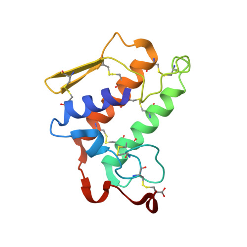
    Phospholipase A2 catalyses the hydrolysis of the ester bond of 3-sn-phosphoglycerides. Here, we report the crystal structures of the free and anisic acid-bound triple mutant (K53,56,120M) of bovine pancreatic phospholipase A2. In the bound triple mutant structure, the small organic molecule p-anisic acid is found in the active site, and one of the carboxylate oxygen atoms is coordinated to the functionally important primary calcium ion. The other carboxylate oxygen atom is hydrogen bonded to the phenolic hydroxyl group of Tyr69. In addition, the bound anisic acid molecule replaces one of the functionally important water molecules in the active site. The residues 60-70, which are in a loop (surface loop), are disordered in most of the bovine pancreatic phospholipase A2 structures. It is interesting to note that these residues are ordered in the bound triple mutant structure but are disordered in the free triple mutant structure. The organic crystallization ingredient 2-methyl-2,4-pentanediol is found near the active site of the free triple mutant structure. The overall tertiary folding and stereochemical parameters for the final models of the free and anisic acid-bound triple mutant are virtually identical.


  • Organizational Affiliation

    Bioinformatics Centre, Indian Institute of Science, Bangalore 560 012, India. sekar@physics.iisc.ernet.in


Macromolecules
Find similar proteins by:  (by identity cutoff)  |  3D Structure
Entity ID: 1
MoleculeChains Sequence LengthOrganismDetailsImage
Phospholipase A2123Bos taurusMutation(s): 3 
EC: 3.1.1.4
UniProt
Find proteins for P00593 (Bos taurus)
Explore P00593 
Go to UniProtKB:  P00593
Entity Groups  
Sequence Clusters30% Identity50% Identity70% Identity90% Identity95% Identity100% Identity
UniProt GroupP00593
Sequence Annotations
Expand
  • Reference Sequence
Small Molecules
Ligands 2 Unique
IDChains Name / Formula / InChI Key2D Diagram3D Interactions
MPD
Query on MPD

Download Ideal Coordinates CCD File 
C [auth A](4S)-2-METHYL-2,4-PENTANEDIOL
C6 H14 O2
SVTBMSDMJJWYQN-YFKPBYRVSA-N
CA
Query on CA

Download Ideal Coordinates CCD File 
B [auth A]CALCIUM ION
Ca
BHPQYMZQTOCNFJ-UHFFFAOYSA-N
Experimental Data & Validation

Experimental Data

  • Method: X-RAY DIFFRACTION
  • Resolution: 1.85 Å
  • R-Value Free: 0.232 
  • R-Value Work: 0.193 
  • Space Group: P 31 2 1
Unit Cell:
Length ( Å )Angle ( ˚ )
a = 46.76α = 90
b = 46.76β = 90
c = 102.71γ = 120
Software Package:
Software NamePurpose
DENZOdata reduction
CNSrefinement
SCALEPACKdata scaling

Structure Validation

View Full Validation Report


Entry History 

Deposition Data

  • Released Date: 2003-07-15 
  • Deposition Author(s): Sekar, K.

Revision History  (Full details and data files)

  • Version 1.0: 2003-07-15
    Type: Initial release
  • Version 1.1: 2008-04-26
    Changes: Version format compliance
  • Version 1.2: 2011-07-13
    Changes: Version format compliance
  • Version 1.3: 2021-10-27
    Changes: Database references, Derived calculations
  • Version 1.4: 2023-12-27
    Changes: Data collection