1O0R

Crystal structure of the catalytic domain of bovine beta1,4-galactosyltransferase complex with UDP-galactose


Experimental Data Snapshot

  • Method: X-RAY DIFFRACTION
  • Resolution: 2.30 Å
  • R-Value Free: 0.251 
  • R-Value Work: 0.203 
  • R-Value Observed: 0.203 

wwPDB Validation   3D Report Full Report


Ligand Structure Quality Assessment 


This is version 1.3 of the entry. See complete history


Literature

CRYSTAL STRUCTURE OF BETA 1,4-GALACTOSYLTRANSFERASE COMPLEX WITH UDP-GAL REVEALS AN OLIGOSACCHARIDE ACCEPTOR BINDING SITE

Ramakrishnan, B.Balaji, P.V.Qasba, P.K.

(2002) J Mol Biol 318: 491-502

  • DOI: https://doi.org/10.1016/S0022-2836(02)00020-7
  • Primary Citation of Related Structures:  
    1O0R

  • PubMed Abstract: 

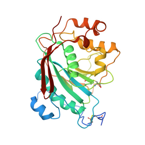
    The crystal structure of the catalytic domain of bovine beta1,4-galactosyltransferase (Gal-T1) co-crystallized with UDP-Gal and MnCl(2) has been solved at 2.8 A resolution. The structure not only identifies galactose, the donor sugar binding site in Gal-T1, but also reveals an oligosaccharide acceptor binding site. The galactose moiety of UDP-Gal is found deep inside the catalytic pocket, interacting with Asp252, Gly292, Gly315, Glu317 and Asp318 residues. Compared to the native crystal structure reported earlier, the present UDP-Gal bound structure exhibits a large conformational change in residues 345-365 and a change in the side-chain orientation of Trp314. Thus, the binding of UDP-Gal induces a conformational change in Gal-T1, which not only creates the acceptor binding pocket for N-acetylglucosamine (GlcNAc) but also establishes the binding site for an extended sugar acceptor. The presence of a binding site that accommodates an extended sugar offers an explanation for the observation that an oligosaccharide with GlcNAc at the non-reducing end serves as a better acceptor than the monosaccharide, GlcNAc. Modeling studies using oligosaccharide acceptors indicate that a pentasaccharide, such as N-glycans with GlcNAc at their non-reducing ends, fits the site best. A sequence comparison of the human Gal-T family members indicates that although the binding site for the GlcNAc residue is highly conserved, the site that binds the extended sugar exhibits large variations. This is an indication that different Gal-T family members prefer different types of glycan acceptors with GlcNAc at their non-reducing ends.


  • Organizational Affiliation

    Structural Glycobiology Section, Laboratory of Experimental and Computational Biology, CCR, NCI, Frederick, MD 21702-1201, USA.


Macromolecules
Find similar proteins by:  (by identity cutoff)  |  3D Structure
Entity ID: 1
MoleculeChains Sequence LengthOrganismDetailsImage
beta-1,4-galactosyltransferase
A, B
286Bos taurusMutation(s): 1 
EC: 2.4.1.90
UniProt
Find proteins for P08037 (Bos taurus)
Explore P08037 
Go to UniProtKB:  P08037
Entity Groups  
Sequence Clusters30% Identity50% Identity70% Identity90% Identity95% Identity100% Identity
UniProt GroupP08037
Sequence Annotations
Expand
  • Reference Sequence
Small Molecules
Ligands 5 Unique
IDChains Name / Formula / InChI Key2D Diagram3D Interactions
GDU
Query on GDU

Download Ideal Coordinates CCD File 
C [auth A],
H [auth B]
GALACTOSE-URIDINE-5'-DIPHOSPHATE
C15 H24 N2 O17 P2
HSCJRCZFDFQWRP-ABVWGUQPSA-N
MES
Query on MES

Download Ideal Coordinates CCD File 
F [auth A]2-(N-MORPHOLINO)-ETHANESULFONIC ACID
C6 H13 N O4 S
SXGZJKUKBWWHRA-UHFFFAOYSA-N
SO4
Query on SO4

Download Ideal Coordinates CCD File 
E [auth A],
J [auth B],
K [auth B],
L [auth B]
SULFATE ION
O4 S
QAOWNCQODCNURD-UHFFFAOYSA-L
DIO
Query on DIO

Download Ideal Coordinates CCD File 
G [auth A],
M [auth B]
1,4-DIETHYLENE DIOXIDE
C4 H8 O2
RYHBNJHYFVUHQT-UHFFFAOYSA-N
MN
Query on MN

Download Ideal Coordinates CCD File 
D [auth A],
I [auth B]
MANGANESE (II) ION
Mn
WAEMQWOKJMHJLA-UHFFFAOYSA-N
Experimental Data & Validation

Experimental Data

  • Method: X-RAY DIFFRACTION
  • Resolution: 2.30 Å
  • R-Value Free: 0.251 
  • R-Value Work: 0.203 
  • R-Value Observed: 0.203 
  • Space Group: P 21 21 21
Unit Cell:
Length ( Å )Angle ( ˚ )
a = 50.356α = 90
b = 91.775β = 90
c = 142.467γ = 90
Software Package:
Software NamePurpose
CNSrefinement
DENZOdata reduction
SCALEPACKdata scaling
AMoREphasing

Structure Validation

View Full Validation Report


Ligand Structure Quality Assessment 


Entry History 

Deposition Data

Revision History  (Full details and data files)

  • Version 1.0: 2003-03-04
    Type: Initial release
  • Version 1.1: 2008-04-29
    Changes: Version format compliance
  • Version 1.2: 2011-07-13
    Changes: Version format compliance
  • Version 1.3: 2021-10-27
    Changes: Database references, Derived calculations