1NPL

MANNOSE-SPECIFIC AGGLUTININ (LECTIN) FROM DAFFODIL (NARCISSUS PSEUDONARCISSUS) BULBS IN COMPLEX WITH MANNOSE-ALPHA1,3-MANNOSE


Experimental Data Snapshot

  • Method: X-RAY DIFFRACTION
  • Resolution: 2.00 Å
  • R-Value Free: 0.267 
  • R-Value Work: 0.187 

wwPDB Validation   3D Report Full Report


This is version 2.0 of the entry. See complete history


Literature

Insights into carbohydrate recognition by Narcissus pseudonarcissus lectin: the crystal structure at 2 A resolution in complex with alpha1-3 mannobiose.

Sauerborn, M.K.Wright, L.M.Reynolds, C.D.Grossmann, J.G.Rizkallah, P.J.

(1999) J Mol Biol 290: 185-199

  • DOI: https://doi.org/10.1006/jmbi.1999.2862
  • Primary Citation of Related Structures:  
    1NPL

  • PubMed Abstract: 

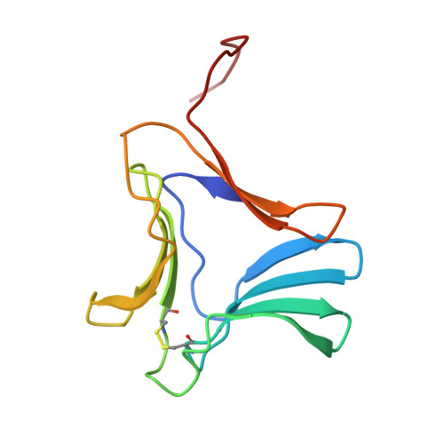
    Carbohydrate recognition by monocot mannose-binding lectins was studied via the crystal structure determination of daffodil (Narcissus pseudonarcissus) lectin. The lectin was extracted from daffodil bulbs, and crystallised in the presence of alpha-1,3 mannobiose. Molecular replacement methods were used to solve the structure using the partially refined model of Hippeastrum hybrid agglutinin as a search model. The structure was refined at 2.0 A resolution to a final R -factor of 18.7 %, and Rfreeof 26.7 %. The main feature of the daffodil lectin structure is the presence of three fully occupied binding pockets per monomer, arranged around the faces of a triangular beta-prism motif. The pockets have identical topology, and can bind mono-, di- or oligosaccharides. Strand exchange forms tightly bound dimers, and higher aggregation states are achieved through hydrophobic patches on the surface, completing a tetramer with internal 222-symmetry. There are therefore 12 fully occupied binding pockets per tetrameric cluster. The tetramer persists in solution, as shown with small-angle X-ray solution scattering. Extensive sideways and out-of-plane interactions between tetramers, some mediated via the ligand, make up the bulk of the lattice contacts.A fourth binding site was also observed. This is unique and has not been observed in similar structures. The site is only partially occupied by a ligand molecule due to the much lower binding affinity. A comparison with the Galanthus nivalis agglutinin/mannopentaose complex suggests an involvement of this site in the recognition mechanism for naturally occurring glycans.


  • Organizational Affiliation

    CCLRC Daresbury Laboratory, Keckwick Lane, Daresbury, Warrington, Cheshire, WA4 4AD, UK.


Macromolecules
Find similar proteins by:  (by identity cutoff)  |  3D Structure
Entity ID: 1
MoleculeChains Sequence LengthOrganismDetailsImage
PROTEIN (AGGLUTININ)109Narcissus pseudonarcissusMutation(s): 0 
UniProt
Find proteins for C0HM45 (Narcissus pseudonarcissus)
Explore C0HM45 
Go to UniProtKB:  C0HM45
Entity Groups  
Sequence Clusters30% Identity50% Identity70% Identity90% Identity95% Identity100% Identity
UniProt GroupC0HM45
Sequence Annotations
Expand
  • Reference Sequence
Oligosaccharides

Help

Entity ID: 2
MoleculeChains Length2D Diagram Glycosylation3D Interactions
alpha-D-mannopyranose-(1-3)-alpha-D-mannopyranose
B, C, D, E
2N/A
Glycosylation Resources
GlyTouCan:  G00891MP
GlyCosmos:  G00891MP
GlyGen:  G00891MP
Small Molecules
Binding Affinity Annotations 
IDSourceBinding Affinity
MAN Binding MOAD:  1NPL Kd: 2.00e+6 (nM) from 1 assay(s)
Experimental Data & Validation

Experimental Data

  • Method: X-RAY DIFFRACTION
  • Resolution: 2.00 Å
  • R-Value Free: 0.267 
  • R-Value Work: 0.187 
  • Space Group: C 2 2 2
Unit Cell:
Length ( Å )Angle ( ˚ )
a = 73.3α = 90
b = 101.2β = 90
c = 37.3γ = 90
Software Package:
Software NamePurpose
MOSFLMdata reduction
SCALAdata scaling
AMoREphasing
REFMACrefinement
CCP4data scaling

Structure Validation

View Full Validation Report


Entry History 

Deposition Data

Revision History  (Full details and data files)

  • Version 1.0: 1998-12-23
    Type: Initial release
  • Version 1.1: 2008-04-27
    Changes: Version format compliance
  • Version 1.2: 2011-07-13
    Changes: Derived calculations, Version format compliance
  • Version 2.0: 2020-07-29
    Type: Remediation
    Reason: Carbohydrate remediation
    Changes: Advisory, Atomic model, Data collection, Database references, Derived calculations, Structure summary