1N8I

Biochemical and Structural Studies of Malate Synthase from Mycobacterium tuberculosis


Experimental Data Snapshot

  • Method: X-RAY DIFFRACTION
  • Resolution: 2.10 Å
  • R-Value Free: 0.231 
  • R-Value Work: 0.185 
  • R-Value Observed: 0.185 

wwPDB Validation   3D Report Full Report


This is version 1.3 of the entry. See complete history


Literature

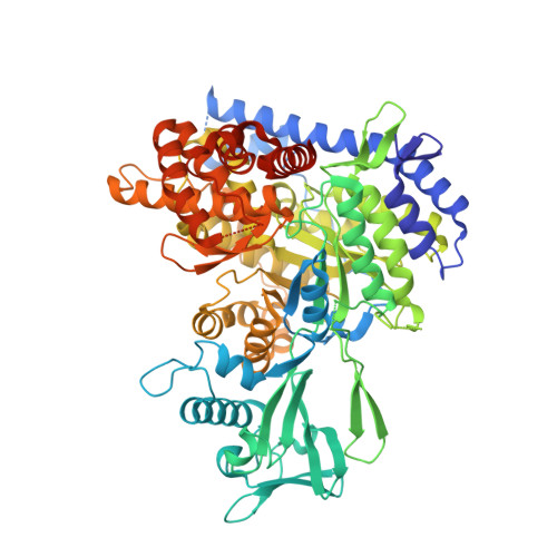
Biochemical and structural studies of malate synthase from Mycobacterium tuberculosis

Smith, C.V.Huang, C.C.Miczak, A.Russell, D.G.Sacchettini, J.C.Honer Zu Bentrup, K.

(2003) J Biol Chem 278: 1735-1743

  • DOI: https://doi.org/10.1074/jbc.M209248200
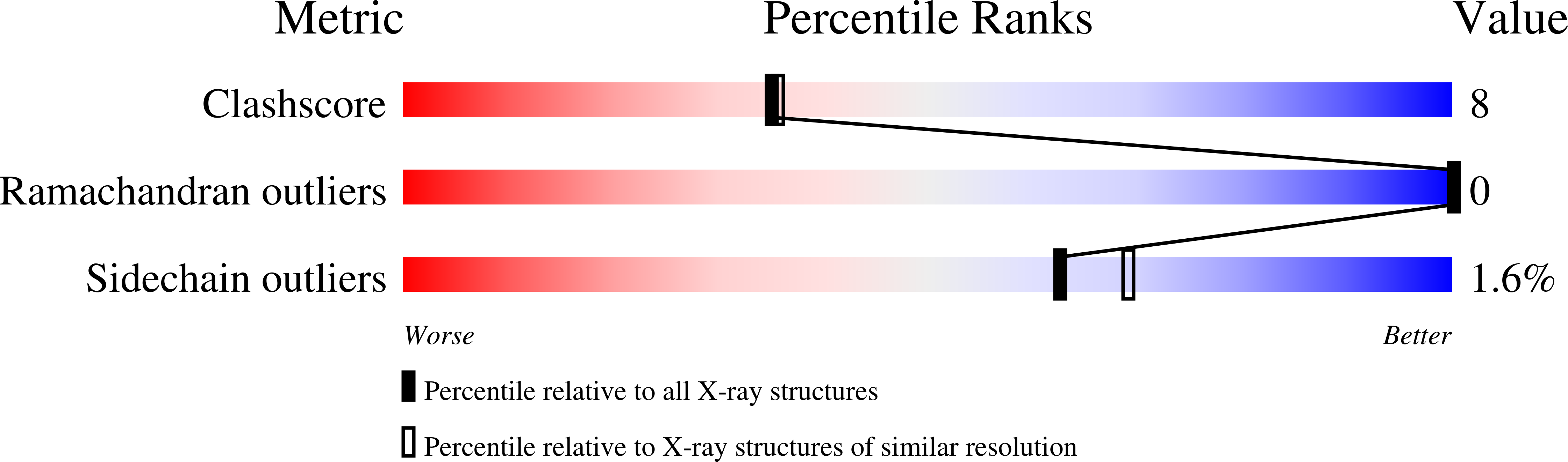
  • Primary Citation of Related Structures:  
    1N8I, 1N8W

  • PubMed Abstract: 

    Establishment or maintenance of a persistent infection by Mycobacterium tuberculosis requires the glyoxylate pathway. This is a bypass of the tricarboxylic acid cycle in which isocitrate lyase and malate synthase (GlcB) catalyze the net incorporation of carbon during growth of microorganisms on acetate or fatty acids as the primary carbon source. The glcB gene from M. tuberculosis, which encodes malate synthase, was cloned, and GlcB was expressed in Escherichia coli. The influence of media conditions on expression in M. tuberculosis indicated that this enzyme is regulated differentially to isocitrate lyase. Purified GlcB had K(m) values of 57 and 30 microm for its substrates glyoxylate and acetyl coenzyme A, respectively, and was inhibited by bromopyruvate, oxalate, and phosphoenolpyruvate. The GlcB structure was solved to 2.1-A resolution in the presence of glyoxylate and magnesium. We also report the structure of GlcB in complex with the products of the reaction, coenzyme A and malate, solved to 2.7-A resolution. Coenzyme A binds in a bent conformation, and the details of its interactions are described, together with implications on the enzyme mechanism.


  • Organizational Affiliation

    Department of Biochemistry and Biophysics, Texas A & M University, College Station, Texas 77843-2128, USA.


Macromolecules
Find similar proteins by:  (by identity cutoff)  |  3D Structure
Entity ID: 1
MoleculeChains Sequence LengthOrganismDetailsImage
Probable malate synthase G741Mycobacterium tuberculosisMutation(s): 0 
Gene Names: GLCB
EC: 4.1.3.2
UniProt
Find proteins for P9WK17 (Mycobacterium tuberculosis (strain ATCC 25618 / H37Rv))
Explore P9WK17 
Go to UniProtKB:  P9WK17
Entity Groups  
Sequence Clusters30% Identity50% Identity70% Identity90% Identity95% Identity100% Identity
UniProt GroupP9WK17
Sequence Annotations
Expand
  • Reference Sequence
Small Molecules
Ligands 2 Unique
IDChains Name / Formula / InChI Key2D Diagram3D Interactions
GLV
Query on GLV

Download Ideal Coordinates CCD File 
C [auth A]GLYOXYLIC ACID
C2 H2 O3
HHLFWLYXYJOTON-UHFFFAOYSA-N
MG
Query on MG

Download Ideal Coordinates CCD File 
B [auth A]MAGNESIUM ION
Mg
JLVVSXFLKOJNIY-UHFFFAOYSA-N
Experimental Data & Validation

Experimental Data

  • Method: X-RAY DIFFRACTION
  • Resolution: 2.10 Å
  • R-Value Free: 0.231 
  • R-Value Work: 0.185 
  • R-Value Observed: 0.185 
  • Space Group: P 43 21 2
Unit Cell:
Length ( Å )Angle ( ˚ )
a = 78.095α = 90
b = 78.095β = 90
c = 223.18γ = 90
Software Package:
Software NamePurpose
HKL-2000data collection
SCALEPACKdata scaling
SOLVEphasing
CNSrefinement
HKL-2000data reduction

Structure Validation

View Full Validation Report


Entry History 

Revision History  (Full details and data files)

  • Version 1.0: 2002-12-18
    Type: Initial release
  • Version 1.1: 2008-04-28
    Changes: Version format compliance
  • Version 1.2: 2011-07-13
    Changes: Version format compliance
  • Version 1.3: 2024-02-14
    Changes: Data collection, Database references, Derived calculations, Structure summary