1N7D

Extracellular domain of the LDL receptor


Experimental Data Snapshot

  • Method: X-RAY DIFFRACTION
  • Resolution: 3.70 Å
  • R-Value Free: 0.382 
  • R-Value Work: 0.381 

wwPDB Validation   3D Report Full Report


This is version 2.1 of the entry. See complete history


Literature

Structure of the LDL receptor extracellular domain at endosomal pH

Rudenko, G.Henry, L.Henderson, K.Ichtchenko, K.Brown, M.S.Goldstein, J.L.Deisenhofer, J.

(2002) Science 298: 2353-2358

  • DOI: https://doi.org/10.1126/science.1078124
  • Primary Citation of Related Structures:  
    1N7D

  • PubMed Abstract: 

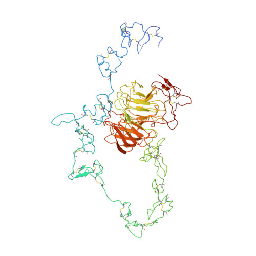
    The low-density lipoprotein receptor mediates cholesterol homeostasis through endocytosis of lipoproteins. It discharges its ligand in the endosome at pH < 6. In the crystal structure at pH = 5.3, the ligand-binding domain (modules R2 to R7) folds back as an arc over the epidermal growth factor precursor homology domain (the modules A, B, beta propeller, and C). The modules R4 and R5, which are critical for lipoprotein binding, associate with the beta propeller via their calcium-binding loop. We propose a mechanism for lipoprotein release in the endosome whereby the beta propeller functions as an alternate substrate for the ligand-binding domain, binding in a calcium-dependent way and promoting lipoprotein release.


  • Organizational Affiliation

    Department of Biochemistry, University of Texas Southwestern Medical Center, 5323 Harry Hines Boulevard Y4-206, Dallas, TX 75390, USA.


Macromolecules
Find similar proteins by:  (by identity cutoff)  |  3D Structure
Entity ID: 1
MoleculeChains Sequence LengthOrganismDetailsImage
Low-density lipoprotein receptor699Homo sapiensMutation(s): 2 
Gene Names: LDLR
UniProt & NIH Common Fund Data Resources
Find proteins for P01130 (Homo sapiens)
Explore P01130 
Go to UniProtKB:  P01130
PHAROS:  P01130
GTEx:  ENSG00000130164 
Entity Groups  
Sequence Clusters30% Identity50% Identity70% Identity90% Identity95% Identity100% Identity
UniProt GroupP01130
Sequence Annotations
Expand
  • Reference Sequence
Oligosaccharides

Help

Entity ID: 2
MoleculeChains Length2D Diagram Glycosylation3D Interactions
alpha-D-mannopyranose-(1-3)-[alpha-D-mannopyranose-(1-6)]beta-D-mannopyranose-(1-4)-2-acetamido-2-deoxy-beta-D-glucopyranose-(1-4)-2-acetamido-2-deoxy-beta-D-glucopyranose
B
5N-Glycosylation
Glycosylation Resources
GlyTouCan:  G22768VO
GlyCosmos:  G22768VO
GlyGen:  G22768VO
Entity ID: 3
MoleculeChains Length2D Diagram Glycosylation3D Interactions
alpha-D-mannopyranose-(1-6)-beta-D-mannopyranose-(1-4)-2-acetamido-2-deoxy-beta-D-glucopyranose-(1-4)-2-acetamido-2-deoxy-beta-D-glucopyranose
C
4N-Glycosylation
Glycosylation Resources
GlyTouCan:  G22573RC
GlyCosmos:  G22573RC
GlyGen:  G22573RC
Experimental Data & Validation

Experimental Data

  • Method: X-RAY DIFFRACTION
  • Resolution: 3.70 Å
  • R-Value Free: 0.382 
  • R-Value Work: 0.381 
  • Space Group: P 31 2 1
Unit Cell:
Length ( Å )Angle ( ˚ )
a = 185.29α = 90
b = 185.29β = 90
c = 85.186γ = 120
Software Package:
Software NamePurpose
MOSFLMdata reduction
SCALAdata scaling
SHARPphasing
CNSrefinement
CCP4data scaling

Structure Validation

View Full Validation Report


Entry History 

Deposition Data

Revision History  (Full details and data files)

  • Version 1.0: 2003-01-21
    Type: Initial release
  • Version 1.1: 2008-04-28
    Changes: Version format compliance
  • Version 1.2: 2011-07-13
    Changes: Non-polymer description, Version format compliance
  • Version 2.0: 2020-07-29
    Type: Remediation
    Reason: Carbohydrate remediation
    Changes: Advisory, Atomic model, Data collection, Derived calculations, Structure summary
  • Version 2.1: 2021-10-27
    Changes: Database references, Structure summary