Solution structure of calmodulin-W-7 complex: the basis of diversity in molecular recognition.
Osawa, M., Swindells, M.B., Tanikawa, J., Tanaka, T., Mase, T., Furuya, T., Ikura, M.(1998) J Mol Biol 276: 165-176
- PubMed: 9514729
- DOI: https://doi.org/10.1006/jmbi.1997.1524
- Primary Citation of Related Structures:
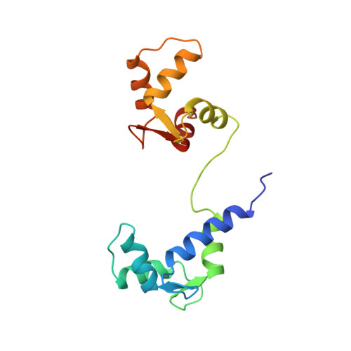
1MUX - PubMed Abstract:
The solution structure of calcium-bound calmodulin (CaM) complexed with an antagonist, N-(6-aminohexyl)-5-chloro-1-naphthalenesulfonamide (W-7), has been determined by multidimensional NMR spectroscopy. The structure consists of one molecule of W-7 binding to each of the two domains of CaM. In each domain, the W-7 chloronaphthalene ring interacts with four methionine methyl groups and other aliphatic or aromatic side-chains in a deep hydrophobic pocket, the site responsible for CaM binding to CaM-dependent enzymes such as myosin light chain kinases (MLCKs) and CaM kinase II. This competitive binding at the same site between W-7 and CaM-dependent enzymes suggests the mechanism by which W-7 inhibits CaM to activate the enzymes. The orientation of the W-7 naphthalene ring in the N-terminal pocket is rotated approximately 40 degrees with respect to that in the C-terminal pocket. The W-7 ring orientation differs significantly from the Trp800 indole ring of smooth muscle MLCK bound to the C-terminal pocket and the phenothiazine ring of trifluoperazine bound to the N or C-terminal pocket. These comparative structural analyses demonstrate that the two hydrophobic pockets of CaM can accommodate a variety of bulky aromatic rings, which provides a plausible structural basis for the diversity in CaM-mediated molecular recognition.
Organizational Affiliation:
Molecular Chemistry Research Chemistry Laboratories, Yamanouchi Pharmaceutical Co., Tsukuba, Japan.