1MT0

ATP-binding domain of hemolysin B from Escherichia coli


Experimental Data Snapshot

  • Method: X-RAY DIFFRACTION
  • Resolution: 2.60 Å
  • R-Value Free: 0.260 
  • R-Value Work: 0.234 
  • R-Value Observed: 0.234 

wwPDB Validation   3D Report Full Report


This is version 1.3 of the entry. See complete history


Literature

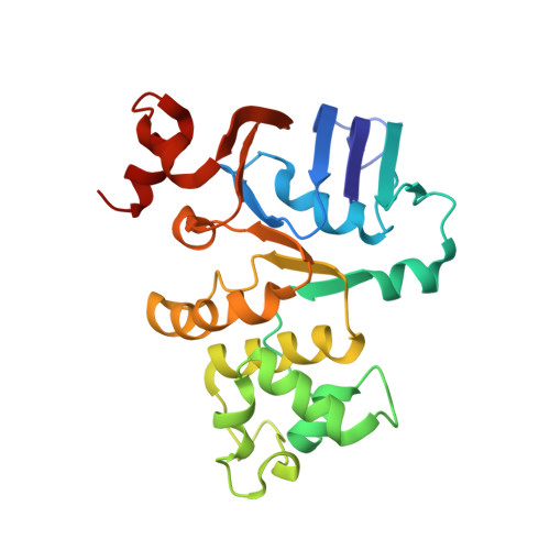
Crystal structure of the nucleotide binding domain of the ABC-transporter hemolysin B: identification of a variable region within ABC helical domains

Schmitt, L.Benabdelhak, H.Blight, M.A.Holland, I.B.Stubbs, M.T.

(2003) J Mol Biol 330: 333-342

  • DOI: https://doi.org/10.1016/s0022-2836(03)00592-8
  • Primary Citation of Related Structures:  
    1MT0

  • PubMed Abstract: 

    The ABC-transporter haemolysin B is a central component of the secretion machinery that translocates the toxin, haemolysin A, in a Sec-independent fashion across both membranes of E. coli. Here, we report the X-ray crystal structure of the nucleotide-binding domain (NBD) of HlyB. The molecule shares the common overall architecture of ABC-transporter NBDs. However, the last three residues of the Walker A motif adopt a 3(10) helical conformation, stabilized by a bound anion. In consequence, this results in an unusual interaction between the Walker A lysine residue and the Walker B glutamate residue. As these residues are normally required to be available for ATP binding, for catalysis and for dimer formation of ABC domains, we suggest that this conformation may represent a latent monomeric form of the NBD. Surprisingly, comparison of available NBD structures revealed a structurally diverse region (SDR) of about 30 residues within the helical arm II domain, unique to each of the eight NBDs analyzed. As this region interacts with the transmembrane part of ABC-transporters, the SDR helps to explain the selectivity and/or targeting of different NBDs to their cognate transmembrane domains.


  • Organizational Affiliation

    Institut für Biochemie, Biozentrum N210, Johann Wolfgang Goethe Universität Frankfurt, Marie-Curie Str. 9, 60439, Frankfurt, Germany. lschmitt@em.uni-frankfurt.de


Macromolecules
Find similar proteins by:  (by identity cutoff)  |  3D Structure
Entity ID: 1
MoleculeChains Sequence LengthOrganismDetailsImage
Hemolysin secretion ATP-binding protein241Escherichia coliMutation(s): 0 
Gene Names: HLYB
UniProt
Find proteins for P08716 (Escherichia coli)
Explore P08716 
Go to UniProtKB:  P08716
Entity Groups  
Sequence Clusters30% Identity50% Identity70% Identity90% Identity95% Identity100% Identity
UniProt GroupP08716
Sequence Annotations
Expand
  • Reference Sequence
Small Molecules
Ligands 1 Unique
IDChains Name / Formula / InChI Key2D Diagram3D Interactions
SO4
Query on SO4

Download Ideal Coordinates CCD File 
B [auth A]SULFATE ION
O4 S
QAOWNCQODCNURD-UHFFFAOYSA-L
Experimental Data & Validation

Experimental Data

  • Method: X-RAY DIFFRACTION
  • Resolution: 2.60 Å
  • R-Value Free: 0.260 
  • R-Value Work: 0.234 
  • R-Value Observed: 0.234 
  • Space Group: P 41 21 2
Unit Cell:
Length ( Å )Angle ( ˚ )
a = 105.2α = 90
b = 105.2β = 90
c = 125.4γ = 90
Software Package:
Software NamePurpose
DENZOdata reduction
SCALEPACKdata scaling
SOLVEphasing
CNSrefinement

Structure Validation

View Full Validation Report


Entry History 

Deposition Data

Revision History  (Full details and data files)

  • Version 1.0: 2003-06-17
    Type: Initial release
  • Version 1.1: 2008-04-28
    Changes: Version format compliance
  • Version 1.2: 2011-07-13
    Changes: Version format compliance
  • Version 1.3: 2024-02-14
    Changes: Data collection, Database references, Derived calculations