1MSM

The HIV protease (mutant Q7K L33I L63I) complexed with KNI-764 (an inhibitor)


Experimental Data Snapshot

  • Method: X-RAY DIFFRACTION
  • Resolution: 2.00 Å
  • R-Value Free: 0.236 
  • R-Value Work: 0.209 

wwPDB Validation   3D Report Full Report


This is version 1.5 of the entry. See complete history


Literature

A structural and thermodynamic escape mechanism from a drug resistant mutation of the HIV-1 protease.

Vega, S.Kang, L.-W.Velazquez-Campoy, A.Kiso, Y.Amzel, L.M.Freire, E.

(2004) Proteins 55: 594-602

  • DOI: https://doi.org/10.1002/prot.20069
  • Primary Citation of Related Structures:  
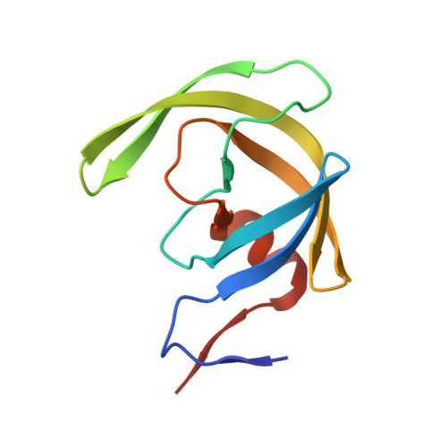
    1MRW, 1MRX, 1MSM, 1MSN

  • PubMed Abstract: 

    The efficacy of HIV-1 protease inhibition therapies is often compromised by the appearance of mutations in the protease molecule that lower the binding affinity of inhibitors while maintaining viable catalytic activity and substrate affinity. The V82F/I84V double mutation is located within the binding site cavity and affects all protease inhibitors in clinical use. KNI-764, a second-generation inhibitor currently under development, maintains significant potency against this mutation by entropically compensating for enthalpic losses, thus minimizing the loss in binding affinity. KNI-577 differs from KNI-764 by a single functional group critical to the inhibitor response to the protease mutation. This single difference changes the response of the two inhibitors to the mutation by one order of magnitude. Accordingly, a structural understanding of the inhibitor response will provide important guidelines for the design of inhibitors that are less susceptible to mutations conveying drug resistance. The structures of the two compounds bound to the wild type and V82F/I84V HIV-1 protease have been determined by X-ray crystallography at 2.0 A resolution. The presence of two asymmetric functional groups, linked by rotatable bonds to the inhibitor scaffold, allows KNI-764 to adapt to the mutated binding site cavity more readily than KNI-577, with a single asymmetric group. Both inhibitors lose about 2.5 kcal/mol in binding enthalpy when facing the drug-resistant mutant protease; however KNI-764 gains binding entropy while KNI-577 loses binding entropy. The gain in binding entropy by KNI-764 accounts for its low susceptibility to the drug-resistant mutation. The heat capacity change associated with binding becomes more negative when KNI-764 binds to the mutant protease, consistent with increased desolvation. With KNI-577, the opposite effect is observed. Structurally, the crystallographic B factors increase for KNI-764 when it is bound to the drug-resistant mutant. The opposite is observed for KNI-577. Consistent with these observations, it appears that KNI-764 is able to gain binding entropy by a two-fold mechanism: it gains solvation entropy by burying itself deeper within the binding pocket and gains conformational entropy by losing interaction with the protease.


  • Organizational Affiliation

    Department of Biology, The Johns Hopkins University, Baltimore, MD 21218, USA.


Macromolecules
Find similar proteins by:  (by identity cutoff)  |  3D Structure
Entity ID: 1
MoleculeChains Sequence LengthOrganismDetailsImage
POL polyprotein
A, B
99Human immunodeficiency virus 1Mutation(s): 3 
EC: 3.4.23.16
UniProt
Find proteins for P03367 (Human immunodeficiency virus type 1 group M subtype B (isolate BRU/LAI))
Explore P03367 
Go to UniProtKB:  P03367
Entity Groups  
Sequence Clusters30% Identity50% Identity70% Identity90% Identity95% Identity100% Identity
UniProt GroupP03367
Sequence Annotations
Expand
  • Reference Sequence
Small Molecules
Ligands 1 Unique
IDChains Name / Formula / InChI Key2D Diagram3D Interactions
JE2
Query on JE2

Download Ideal Coordinates CCD File 
C [auth B](4R)-3-{(2S,3S)-2-hydroxy-3-[(3-hydroxy-2-methylbenzoyl)amino]-4-phenylbutanoyl}-5,5-dimethyl-N-(2-methylbenzyl)-1,3-thiazolidine-4-carboxamide
C32 H37 N3 O5 S
CUFQBQOBLVLKRF-RZDMPUFOSA-N
Binding Affinity Annotations 
IDSourceBinding Affinity
JE2 BindingDB:  1MSM Ki: min: 0.02, max: 35 (nM) from 17 assay(s)
EC50: 2730 (nM) from 1 assay(s)
-TΔS: min: -1.02e+2, max: -2.64e+1 (kJ/mol) from 35 assay(s)
ΔH: min: -3.34e+1, max: 73.15 (kJ/mol) from 36 assay(s)
ΔG: min: -6.23e+1, max: -2.88e+1 (kJ/mol) from 36 assay(s)
Binding MOAD:  1MSM Kd: 0.03 (nM) from 1 assay(s)
K64 PDBBind:  1MSM Kd: 0.03 (nM) from 1 assay(s)
Biologically Interesting Molecules (External Reference) 1 Unique
Entity ID: 2
IDChains NameType/Class2D Diagram3D Interactions
PRD_000566 (JE2)
Query on PRD_000566
C [auth B]KNI-764Peptide-like / Enzyme inhibitor
Experimental Data & Validation

Experimental Data

  • Method: X-RAY DIFFRACTION
  • Resolution: 2.00 Å
  • R-Value Free: 0.236 
  • R-Value Work: 0.209 
  • Space Group: P 21 21 2
Unit Cell:
Length ( Å )Angle ( ˚ )
a = 57.67α = 90
b = 85.819β = 90
c = 46.528γ = 90
Software Package:
Software NamePurpose
DENZOdata reduction
SCALEPACKdata scaling
CNSrefinement
CNSphasing

Structure Validation

View Full Validation Report


Entry History 

Deposition Data

Revision History  (Full details and data files)

  • Version 1.0: 2003-11-04
    Type: Initial release
  • Version 1.1: 2008-04-28
    Changes: Version format compliance
  • Version 1.2: 2011-07-13
    Changes: Atomic model, Database references, Derived calculations, Non-polymer description, Structure summary, Version format compliance
  • Version 1.3: 2012-12-12
    Changes: Other
  • Version 1.4: 2021-10-27
    Changes: Database references, Derived calculations, Structure summary
  • Version 1.5: 2024-02-14
    Changes: Data collection, Refinement description