X-ray Structure of a Maquette Scaffold
Huang, S.S., Gibney, B.R., Stayrook, S.E., Dutton, P.L., Lewis, M.(2003) J Mol Biol 326: 1219-1225
- PubMed: 12589764
- DOI: https://doi.org/10.1016/s0022-2836(02)01441-9
- Primary Citation of Related Structures:
1M3W - PubMed Abstract:
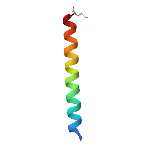
Maquettes are de novo designed mimicries of nature used to test the construction and engineering criteria of oxidoreductases. One type of scaffold used in maquette construction is a four-alpha-helical bundle. The sequence of the four-alpha-helix bundle maquettes follows a heptad repeat pattern typical of left-handed coiled-coils. Initial designs were molten globular due partly to the minimalist approach taken by the designers. Subsequent iterative redesign generated several structured scaffolds with similar heme binding properties. Variant [I(6)F(13)](2), a structured scaffold, was partially resolved with NMR spectroscopy and found to have a set of mobile inter-helical packing interfaces. Here, the X-ray structure of a similar peptide ([I(6)F(13)M(31)](2) i.e. ([CGGG EIWKL HEEFLKK FEELLKL HEERLKKM](2))(2) which we call L31M), has been solved using MAD phasing and refined to 2.8A resolution. The structure shows that the maquette scaffold is an anti-parallel four-helix bundle with "up-up-down-down" topology. No pre-formed heme-binding pocket exists in the protein scaffold. We report unexpected inter-helical crossing angles, residue positions and translations between the helices. The crossing angles between the parallel helices are -5 degrees rather than the expected +20 degrees for typical left-handed coiled-coils. Deviation of the scaffold from the design is likely due to the distribution and size of hydrophobic residues. The structure of L31M points out that four identical helices may interact differently in a bundle and heptad repeats with an alternating [HPPHHPP]/[HPPHHPH] (H: hydrophobic, P: polar) pattern are not a sufficient design criterion to generate left-hand coiled-coils.
Organizational Affiliation:
The Johnson Research Foundation, Department of Biochemistry and Biophysics, School of Medicine, University of Pennsylvania, 37th and Hamilton Walk, Philadelphia, PA 19102-6059, USA.