1LY8

The crystal structure of a mutant enzyme of Coprinus cinereus peroxidase provides an understanding of its increased thermostability and insight into modelling of protein structures


Experimental Data Snapshot

  • Method: X-RAY DIFFRACTION
  • Resolution: 2.05 Å
  • R-Value Free: 0.249 
  • R-Value Work: 0.202 
  • R-Value Observed: 0.202 

wwPDB Validation   3D Report Full Report


Ligand Structure Quality Assessment 


This is version 1.6 of the entry. See complete history


Literature

The structure of a mutant enzyme of Coprinus cinereus peroxidase provides an understanding of its increased thermostability.

Houborg, K.Harris, P.Poulsen, J.C.Schneider, P.Svendsen, A.Larsen, S.

(2003) Acta Crystallogr D Biol Crystallogr 59: 997-1003

  • DOI: https://doi.org/10.1107/s0907444903006784
  • Primary Citation of Related Structures:  
    1LY8

  • PubMed Abstract: 

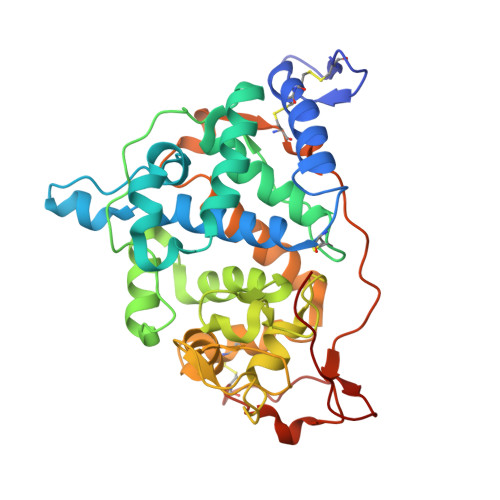
    Seven amino-acid substitutions introduced into the 343 amino-acid-long sequence of Coprinus cinereus peroxidase (CiP) led to a mutant enzyme (TS-rCiP) which is more stable than the native enzyme at higher temperature, pH and hydrogen peroxide concentrations. It is therefore more suitable for industrial applications. A structure determination was conducted on a deglycosylated but still active form of TS-rCiP based on X-ray diffraction data to 2.05 A resolution measured on a crystal cooled to 100 K and refined to R = 0.202 and R(free) = 0.249. The increased stability of the TS-rCiP enzyme can be understood from the structural changes of the TS-rCiP structure revealed by a comparative analysis with other known CiP structures. One of the more significant changes caused by three of the substitutions, I49S, V53A and T121A, is the conversion of a hydrophobic pocket into a hydrophilic pocket with associated changes in the water structure and the hydrogen-bonding interactions. The E239G substitution, which gives rise to increased thermostability at high pH, creates changes in the water structure and in the orientation of a phenylalanine (Phe236) in its vicinity. The three substitutions M166F, M242 and Y242F introduced to increase the oxidative stability do not introduce any structural changes.


  • Organizational Affiliation

    Centre for Crystallographic Studies, University of Copenhagen, Universitetsparken 5, 2100 Copenhagen, Denmark.


Macromolecules
Find similar proteins by:  (by identity cutoff)  |  3D Structure
Entity ID: 1
MoleculeChains Sequence LengthOrganismDetailsImage
Peroxidase
A, B
343Coprinopsis cinereaMutation(s): 7 
Gene Names: CIP1
EC: 1.11.1.7
UniProt
Find proteins for P28314 (Coprinopsis cinerea)
Explore P28314 
Go to UniProtKB:  P28314
Entity Groups  
Sequence Clusters30% Identity50% Identity70% Identity90% Identity95% Identity100% Identity
UniProt GroupP28314
Sequence Annotations
Expand
  • Reference Sequence
Small Molecules
Ligands 6 Unique
IDChains Name / Formula / InChI Key2D Diagram3D Interactions
HEM
Query on HEM

Download Ideal Coordinates CCD File 
H [auth A],
R [auth B]
PROTOPORPHYRIN IX CONTAINING FE
C34 H32 Fe N4 O4
KABFMIBPWCXCRK-RGGAHWMASA-L
NAG
Query on NAG

Download Ideal Coordinates CCD File 
C [auth A],
M [auth B]
2-acetamido-2-deoxy-beta-D-glucopyranose
C8 H15 N O6
OVRNDRQMDRJTHS-FMDGEEDCSA-N
MAN
Query on MAN

Download Ideal Coordinates CCD File 
E [auth A],
O [auth B]
alpha-D-mannopyranose
C6 H12 O6
WQZGKKKJIJFFOK-PQMKYFCFSA-N
BMA
Query on BMA

Download Ideal Coordinates CCD File 
D [auth A],
N [auth B]
beta-D-mannopyranose
C6 H12 O6
WQZGKKKJIJFFOK-RWOPYEJCSA-N
GOL
Query on GOL

Download Ideal Coordinates CCD File 
I [auth A]
J [auth A]
K [auth A]
L [auth A]
S [auth B]
I [auth A],
J [auth A],
K [auth A],
L [auth A],
S [auth B],
T [auth B],
U [auth B],
V [auth B],
W [auth B],
X [auth B],
Y [auth B],
Z [auth B]
GLYCEROL
C3 H8 O3
PEDCQBHIVMGVHV-UHFFFAOYSA-N
CA
Query on CA

Download Ideal Coordinates CCD File 
F [auth A],
G [auth A],
P [auth B],
Q [auth B]
CALCIUM ION
Ca
BHPQYMZQTOCNFJ-UHFFFAOYSA-N
Experimental Data & Validation

Experimental Data

  • Method: X-RAY DIFFRACTION
  • Resolution: 2.05 Å
  • R-Value Free: 0.249 
  • R-Value Work: 0.202 
  • R-Value Observed: 0.202 
  • Space Group: P 21 21 2
Unit Cell:
Length ( Å )Angle ( ˚ )
a = 74.49α = 90
b = 114.86β = 90
c = 73.61γ = 90
Software Package:
Software NamePurpose
DENZOdata reduction
SCALEPACKdata scaling
AMoREphasing
CNSrefinement

Structure Validation

View Full Validation Report


Ligand Structure Quality Assessment 


Entry History 

Deposition Data

Revision History  (Full details and data files)

  • Version 1.0: 2002-06-14
    Type: Initial release
  • Version 1.1: 2008-04-28
    Changes: Version format compliance
  • Version 1.2: 2011-07-13
    Changes: Non-polymer description, Version format compliance
  • Version 1.3: 2011-11-16
    Changes: Atomic model
  • Version 1.4: 2018-03-07
    Changes: Data collection
  • Version 1.5: 2020-07-29
    Type: Remediation
    Reason: Carbohydrate remediation
    Changes: Data collection, Derived calculations, Structure summary
  • Version 1.6: 2021-10-27
    Changes: Database references, Structure summary