1LO9

X-ray crystal structure of 4-hydroxybenzoyl CoA thioesterase mutant D17N complexed with 4-hydroxybenzoyl CoA


Experimental Data Snapshot

  • Method: X-RAY DIFFRACTION
  • Resolution: 2.80 Å
  • R-Value Free: 0.247 
  • R-Value Work: 0.147 
  • R-Value Observed: 0.148 

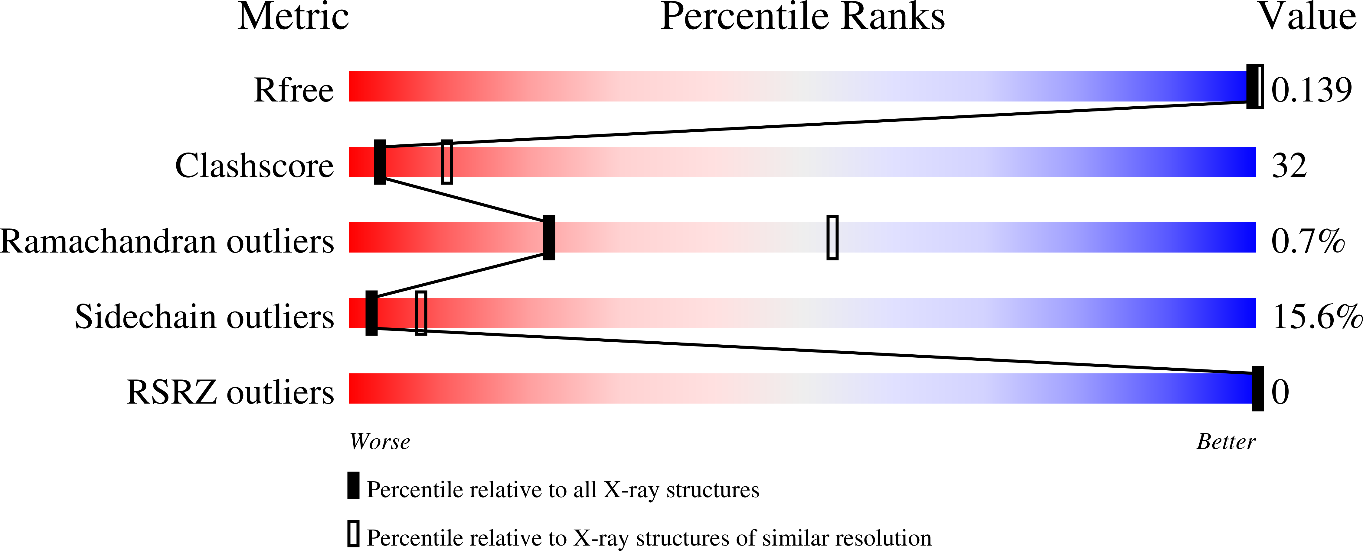
wwPDB Validation   3D Report Full Report


Ligand Structure Quality Assessment 


This is version 1.5 of the entry. See complete history


Literature

X-ray crystallographic analyses of inhibitor and substrate complexes of wild-type and mutant 4-hydroxybenzoyl-CoA thioesterase.

Thoden, J.B.Holden, H.M.Zhuang, Z.Dunaway-Mariano, D.

(2002) J Biol Chem 277: 27468-27476

  • DOI: https://doi.org/10.1074/jbc.M203904200
  • Primary Citation of Related Structures:  
    1LO7, 1LO8, 1LO9

  • PubMed Abstract: 

    The metabolic pathway by which 4-chlorobenzoate is degraded to 4-hydroxybenzoate in the soil-dwelling microbe Pseudomonas sp. strain CBS-3 consists of three enzymes including 4-hydroxybenzoyl-CoA thioesterase. The structure of the unbound form of this thioesterase has been shown to contain the so-called "hot dog" fold with a large helix packed against a five-stranded anti-parallel beta-sheet. To address the manner in which the enzyme accommodates the substrate within the active site, two inhibitors have been synthesized, namely 4-hydroxyphenacyl-CoA and 4-hydroxybenzyl-CoA. Here we describe the structural analyses of the enzyme complexed with these two inhibitors determined and refined to 1.5 and 1.8 A resolution, respectively. These studies indicate that only one protein side chain, Ser(91), participates directly in ligand binding. All of the other interactions between the protein and the inhibitors are mediated through backbone peptidic NH groups, carbonyl oxygens, and/or solvents. The structures of the enzyme-inhibitor complexes suggest that both a hydrogen bond and the positive end of a helix dipole moment serve to polarize the electrons away from the carbonyl carbon of the acyl group, thereby making it more susceptible to nucleophilic attack. Additionally, these studies demonstrate that the carboxylate group of Asp(17) is approximately 3.2 A from the carbonyl carbon of the acyl group. To address the role of Asp(17), the structure of the site-directed mutant protein D17N with bound substrate has also been determined. Taken together, these investigations suggest that the reaction mechanism may proceed through an acyl enzyme intermediate.


  • Organizational Affiliation

    Department of Biochemistry, University of Wisconsin, 433 Babcock Drive, Madison, WI 53706-1544, USA.


Macromolecules
Find similar proteins by:  (by identity cutoff)  |  3D Structure
Entity ID: 1
MoleculeChains Sequence LengthOrganismDetailsImage
4-hydroxybenzoyl-CoA Thioesterase141Pseudomonas sp. CBS3Mutation(s): 1 
Gene Names: 4HBT_PSESP
EC: 3.1.2.23
UniProt
Find proteins for P56653 (Pseudomonas sp. (strain CBS-3))
Explore P56653 
Go to UniProtKB:  P56653
Entity Groups  
Sequence Clusters30% Identity50% Identity70% Identity90% Identity95% Identity100% Identity
UniProt GroupP56653
Sequence Annotations
Expand
  • Reference Sequence
Small Molecules
Ligands 1 Unique
IDChains Name / Formula / InChI Key2D Diagram3D Interactions
BCA
Query on BCA

Download Ideal Coordinates CCD File 
B [auth A]4-HYDROXYBENZOYL COENZYME A
C28 H40 N7 O18 P3 S
LTVXPVBFJBTNIJ-TYHXJLICSA-N
Experimental Data & Validation

Experimental Data

  • Method: X-RAY DIFFRACTION
  • Resolution: 2.80 Å
  • R-Value Free: 0.247 
  • R-Value Work: 0.147 
  • R-Value Observed: 0.148 
  • Space Group: I 2 2 2
Unit Cell:
Length ( Å )Angle ( ˚ )
a = 52.5α = 90
b = 55.8β = 90
c = 93.7γ = 90
Software Package:
Software NamePurpose
AMoREphasing
TNTrefinement
FRAMBOdata collection
SAINTdata scaling

Structure Validation

View Full Validation Report


Ligand Structure Quality Assessment 


Entry History 

Deposition Data

Revision History  (Full details and data files)

  • Version 1.0: 2002-05-13
    Type: Initial release
  • Version 1.1: 2008-04-28
    Changes: Version format compliance
  • Version 1.2: 2011-07-13
    Changes: Version format compliance
  • Version 1.3: 2017-10-11
    Changes: Refinement description
  • Version 1.4: 2021-10-27
    Changes: Database references, Derived calculations
  • Version 1.5: 2023-08-16
    Changes: Data collection, Refinement description