1KXP

CRYSTAL STRUCTURE OF HUMAN VITAMIN D-BINDING PROTEIN IN COMPLEX WITH SKELETAL ACTIN


Experimental Data Snapshot

  • Method: X-RAY DIFFRACTION
  • Resolution: 2.10 Å
  • R-Value Free: 0.236 
  • R-Value Work: 0.217 

wwPDB Validation   3D Report Full Report


This is version 1.3 of the entry. See complete history


Literature

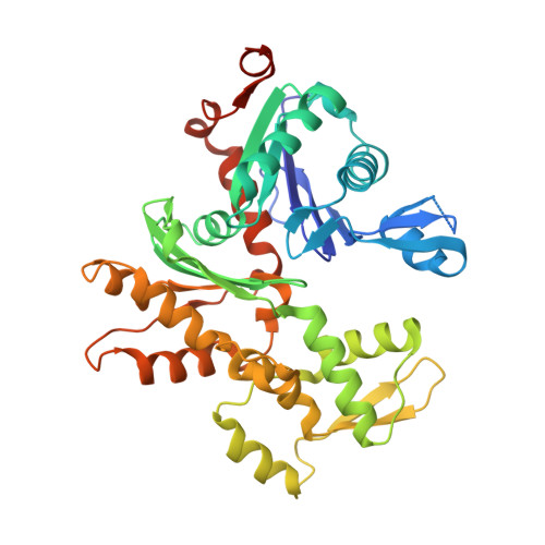
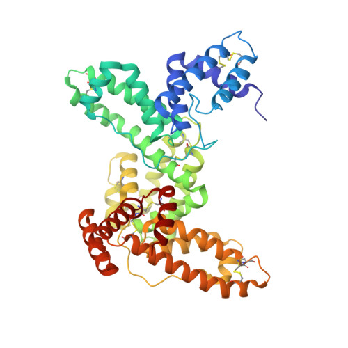
Crystal structures of the vitamin D-binding protein and its complex with actin: structural basis of the actin-scavenger system.

Otterbein, L.R.Cosio, C.Graceffa, P.Dominguez, R.

(2002) Proc Natl Acad Sci U S A 99: 8003-8008

  • DOI: https://doi.org/10.1073/pnas.122126299
  • Primary Citation of Related Structures:  
    1KW2, 1KXP

  • PubMed Abstract: 

    Actin is the most abundant protein in eukaryotic cells, but its release from cells into blood vessels can be lethal, being associated with clinical situations including hepatic necrosis and septic shock. A homeostatic mechanism, termed the actin-scavenger system, is responsible for the depolymerization and removal of actin from the circulation. During the first phase of this mechanism, gelsolin severs the actin filaments. In the second phase, the vitamin D-binding protein (DBP) traps the actin monomers, which accelerates their clearance. We have determined the crystal structures of DBP by itself and complexed with actin to 2.1 A resolution. Similar to its homologue serum albumin, DBP consists of three related domains. Yet, in DBP a strikingly different organization of the domains gives rise to a large actin-binding cavity. After complex formation the three domains of DBP move slightly to "clamp" onto actin subdomain 3 and to a lesser extent subdomain 1. Contacts between actin and DBP throughout their extensive 3,454-A(2) intermolecular interface involve a mixture of hydrophobic, electrostatic, and solvent-mediated interactions. The area of actin covered by DBP within the complex approximately equals the sum of those covered by gelsolin and profilin. Moreover, certain interactions of DBP with actin mirror those observed in the actin-gelsolin complex, which may explain how DBP can compete effectively with gelsolin for actin binding. Formation of the strong actin-DBP complex proceeds with limited conformational changes to both proteins, demonstrating how DBP has evolved to become an effective actin-scavenger protein.


  • Organizational Affiliation

    Boston Biomedical Research Institute, 64 Grove Street, Watertown, MA 02472, USA.


Macromolecules
Find similar proteins by:  (by identity cutoff)  |  3D Structure
Entity ID: 1
MoleculeChains Sequence LengthOrganismDetailsImage
ACTIN,ALPHA SKELETAL MUSCLE375Oryctolagus cuniculusMutation(s): 0 
UniProt
Find proteins for P68135 (Oryctolagus cuniculus)
Explore P68135 
Go to UniProtKB:  P68135
Entity Groups  
Sequence Clusters30% Identity50% Identity70% Identity90% Identity95% Identity100% Identity
UniProt GroupP68135
Sequence Annotations
Expand
  • Reference Sequence
Find similar proteins by:  (by identity cutoff)  |  3D Structure
Entity ID: 2
MoleculeChains Sequence LengthOrganismDetailsImage
HUMAN VITAMIN D-BINDING PROTEINB [auth D]458Homo sapiensMutation(s): 0 
UniProt & NIH Common Fund Data Resources
Find proteins for P02774 (Homo sapiens)
Explore P02774 
Go to UniProtKB:  P02774
PHAROS:  P02774
GTEx:  ENSG00000145321 
Entity Groups  
Sequence Clusters30% Identity50% Identity70% Identity90% Identity95% Identity100% Identity
UniProt GroupP02774
Sequence Annotations
Expand
  • Reference Sequence
Small Molecules
Ligands 2 Unique
IDChains Name / Formula / InChI Key2D Diagram3D Interactions
ATP
Query on ATP

Download Ideal Coordinates CCD File 
D [auth A]ADENOSINE-5'-TRIPHOSPHATE
C10 H16 N5 O13 P3
ZKHQWZAMYRWXGA-KQYNXXCUSA-N
MG
Query on MG

Download Ideal Coordinates CCD File 
C [auth A]MAGNESIUM ION
Mg
JLVVSXFLKOJNIY-UHFFFAOYSA-N
Experimental Data & Validation

Experimental Data

  • Method: X-RAY DIFFRACTION
  • Resolution: 2.10 Å
  • R-Value Free: 0.236 
  • R-Value Work: 0.217 
  • Space Group: P 21 21 21
Unit Cell:
Length ( Å )Angle ( ˚ )
a = 80.758α = 90
b = 87.364β = 90
c = 162.686γ = 90
Software Package:
Software NamePurpose
DENZOdata reduction
SCALEPACKdata scaling
CNSrefinement
CNSphasing

Structure Validation

View Full Validation Report


Entry History 

Deposition Data

Revision History  (Full details and data files)

  • Version 1.0: 2002-06-19
    Type: Initial release
  • Version 1.1: 2008-04-27
    Changes: Version format compliance
  • Version 1.2: 2011-07-13
    Changes: Derived calculations, Version format compliance
  • Version 1.3: 2013-09-18
    Changes: Derived calculations