1JZM

Crystal Structure of Scapharca inaequivalvis HbI, I114M Mutant in the Absence of ligand.


Experimental Data Snapshot

  • Method: X-RAY DIFFRACTION
  • Resolution: 1.90 Å
  • R-Value Free: 0.201 
  • R-Value Work: 0.171 
  • R-Value Observed: 0.171 

wwPDB Validation   3D Report Full Report


Ligand Structure Quality Assessment 


This is version 1.4 of the entry. See complete history


Literature

Restricting the Ligand-Linked Heme Movement in Scapharca Dimeric Hemoglobin Reveals Tight Coupling between Distal and Proximal Contributions to Cooperativity.

Knapp, J.E.Gibson, Q.H.Cushing, L.Royer Jr., W.E.

(2001) Biochemistry 40: 14795-14805

  • DOI: https://doi.org/10.1021/bi011071t
  • Primary Citation of Related Structures:  
    1JWN, 1JZK, 1JZL, 1JZM

  • PubMed Abstract: 

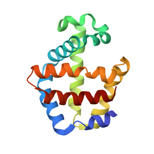
    Cooperative ligand binding in the dimeric hemoglobin from the blood clam Scapharca inaequivalvis results primarily from tertiary, rather than quaternary, structural changes. Ligand binding is coupled with conformational changes of key residues, including Phe 97, which is extruded from the proximal heme pocket, and the heme group, which moves deeper into the heme pocket. We have tested the role of the heme movement in cooperative function by mutating Ile 114, at the base of the heme pocket. Replacement of this residue with a Met did not disturb the hemoglobin structure or significantly alter equilibrium ligand binding properties. In contrast, substitution with a Phe at position 114 inhibits the ligand-linked movement of the heme group, and substantially reduces oxygen affinity and cooperativity. As the extent of heme movement to the normal position of the ligated state is diminished, Phe 97 is inhibited from its movement into the interface upon ligand binding. These results indicate a tight coupling between these two key cooperative transitions and suggest that the heme movement may be an obligatory trigger for expulsion of Phe 97 from the heme pocket.


  • Organizational Affiliation

    Department of Biochemistry and Molecular Pharmacology, University of Massachusetts Medical School, Worcester, Massachusetts 01655, USA.


Macromolecules
Find similar proteins by:  (by identity cutoff)  |  3D Structure
Entity ID: 1
MoleculeChains Sequence LengthOrganismDetailsImage
GLOBIN I - ARK SHELL
A, B
146Anadara inaequivalvisMutation(s): 1 
UniProt
Find proteins for P02213 (Anadara inaequivalvis)
Explore P02213 
Go to UniProtKB:  P02213
Entity Groups  
Sequence Clusters30% Identity50% Identity70% Identity90% Identity95% Identity100% Identity
UniProt GroupP02213
Sequence Annotations
Expand
  • Reference Sequence
Small Molecules
Ligands 1 Unique
IDChains Name / Formula / InChI Key2D Diagram3D Interactions
HEM
Query on HEM

Download Ideal Coordinates CCD File 
C [auth A],
D [auth B]
PROTOPORPHYRIN IX CONTAINING FE
C34 H32 Fe N4 O4
KABFMIBPWCXCRK-RGGAHWMASA-L
Experimental Data & Validation

Experimental Data

  • Method: X-RAY DIFFRACTION
  • Resolution: 1.90 Å
  • R-Value Free: 0.201 
  • R-Value Work: 0.171 
  • R-Value Observed: 0.171 
  • Space Group: C 2 2 21
Unit Cell:
Length ( Å )Angle ( ˚ )
a = 91.82α = 90
b = 44.49β = 90
c = 143.93γ = 90
Software Package:
Software NamePurpose
CNSrefinement
DENZOdata reduction
SCALEPACKdata scaling
CNSphasing

Structure Validation

View Full Validation Report


Ligand Structure Quality Assessment 


Entry History 

Deposition Data

Revision History  (Full details and data files)

  • Version 1.0: 2001-12-19
    Type: Initial release
  • Version 1.1: 2008-04-27
    Changes: Version format compliance
  • Version 1.2: 2011-07-13
    Changes: Version format compliance
  • Version 1.3: 2021-10-27
    Changes: Database references, Derived calculations
  • Version 1.4: 2023-08-16
    Changes: Data collection, Refinement description