1JUQ

GGA3 VHS domain complexed with C-terminal peptide from cation-dependent Mannose 6-phosphate receptor


Experimental Data Snapshot

  • Method: X-RAY DIFFRACTION
  • Resolution: 2.20 Å
  • R-Value Free: 0.254 
  • R-Value Work: 0.221 

wwPDB Validation   3D Report Full Report


This is version 1.3 of the entry. See complete history


Literature

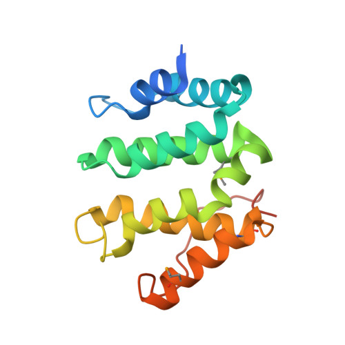
Structural basis for acidic-cluster-dileucine sorting-signal recognition by VHS domains.

Misra, S.Puertollano, R.Kato, Y.Bonifacino, J.S.Hurley, J.H.

(2002) Nature 415: 933-937

  • DOI: https://doi.org/10.1038/415933a
  • Primary Citation of Related Structures:  
    1JPL, 1JUQ

  • PubMed Abstract: 

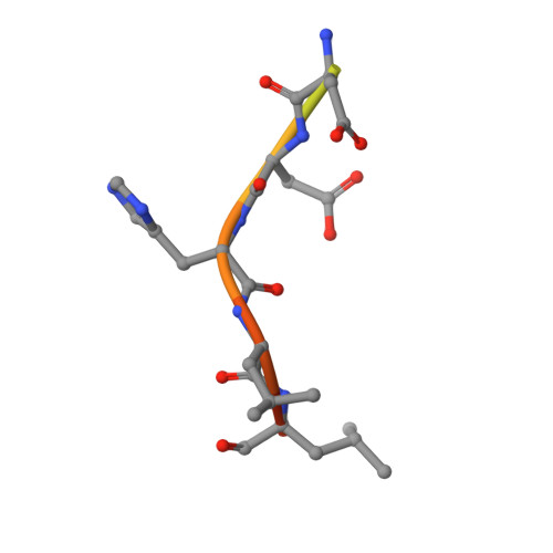
    Specific sorting signals direct transmembrane proteins to the compartments of the endosomal-lysosomal system. Acidic-cluster-dileucine signals present within the cytoplasmic tails of sorting receptors, such as the cation-independent and cation-dependent mannose-6-phosphate receptors, are recognized by the GGA (Golgi-localized, gamma-ear-containing, ADP-ribosylation-factor-binding) proteins. The VHS (Vps27p, Hrs and STAM) domains of the GGA proteins are responsible for the highly specific recognition of these acidic-cluster-dileucine signals. Here we report the structures of the VHS domain of human GGA3 complexed with signals from both mannose-6-phosphate receptors. The signals bind in an extended conformation to helices 6 and 8 of the VHS domain. The structures highlight an Asp residue separated by two residues from a dileucine sequence as critical recognition elements. The side chains of the Asp-X-X-Leu-Leu sequence interact with subsites consisting of one electropositive and two shallow hydrophobic pockets, respectively. The rigid spatial alignment of the three binding subsites leads to high specificity.


  • Organizational Affiliation

    Laboratory of Molecular Biology, National Institute of Diabetes and Digestive and Kidney Diseases, National Institutes of Health, Bethesda, Maryland 20892, USA.


Macromolecules
Find similar proteins by:  (by identity cutoff)  |  3D Structure
Entity ID: 1
MoleculeChains Sequence LengthOrganismDetailsImage
ADP-RIBOSYLATION FACTOR BINDING PROTEIN GGA3A,
C [auth B],
E [auth C],
G [auth D]
171Homo sapiensMutation(s): 4 
Gene Names: GGA3
UniProt & NIH Common Fund Data Resources
Find proteins for Q9NZ52 (Homo sapiens)
Explore Q9NZ52 
Go to UniProtKB:  Q9NZ52
PHAROS:  Q9NZ52
GTEx:  ENSG00000125447 
Entity Groups  
Sequence Clusters30% Identity50% Identity70% Identity90% Identity95% Identity100% Identity
UniProt GroupQ9NZ52
Sequence Annotations
Expand
  • Reference Sequence

Find similar proteins by:  Sequence   |   3D Structure  

Entity ID: 2
MoleculeChains Sequence LengthOrganismDetailsImage
Cation-dependent mannose-6-phosphate receptorB [auth E],
D [auth F],
F [auth G],
H
13N/AMutation(s): 0 
UniProt & NIH Common Fund Data Resources
Find proteins for P20645 (Homo sapiens)
Explore P20645 
Go to UniProtKB:  P20645
PHAROS:  P20645
GTEx:  ENSG00000003056 
Entity Groups  
Sequence Clusters30% Identity50% Identity70% Identity90% Identity95% Identity100% Identity
UniProt GroupP20645
Sequence Annotations
Expand
  • Reference Sequence
Small Molecules
Modified Residues  1 Unique
IDChains TypeFormula2D DiagramParent
MSE
Query on MSE
A,
C [auth B],
E [auth C],
G [auth D]
L-PEPTIDE LINKINGC5 H11 N O2 SeMET
Experimental Data & Validation

Experimental Data

  • Method: X-RAY DIFFRACTION
  • Resolution: 2.20 Å
  • R-Value Free: 0.254 
  • R-Value Work: 0.221 
  • Space Group: C 2 2 21
Unit Cell:
Length ( Å )Angle ( ˚ )
a = 123.64α = 90
b = 131.35β = 90
c = 108.14γ = 90
Software Package:
Software NamePurpose
SOLVEphasing
CNSrefinement
DENZOdata reduction
SCALEPACKdata scaling

Structure Validation

View Full Validation Report


Entry History 

Deposition Data

Revision History  (Full details and data files)

  • Version 1.0: 2002-02-27
    Type: Initial release
  • Version 1.1: 2008-04-27
    Changes: Version format compliance
  • Version 1.2: 2011-07-13
    Changes: Version format compliance
  • Version 1.3: 2016-10-12
    Changes: Advisory