1JL9

Crystal Structure of Human Epidermal Growth Factor


Experimental Data Snapshot

  • Method: X-RAY DIFFRACTION
  • Resolution: 3.00 Å
  • R-Value Free: 0.283 
  • R-Value Work: 0.231 

wwPDB Validation   3D Report Full Report


This is version 1.2 of the entry. See complete history


Literature

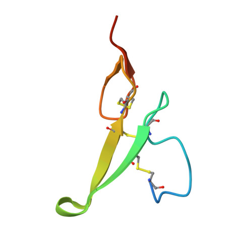
Crystal structure of human epidermal growth factor and its dimerization

Lu, H.S.Chai, J.J.Li, M.Huang, B.R.He, C.H.Bi, R.C.

(2001) J Biol Chem 276: 34913-34917

  • DOI: https://doi.org/10.1074/jbc.M102874200
  • Primary Citation of Related Structures:  
    1JL9

  • PubMed Abstract: 

    Epidermal growth factor (EGF) is a typical growth-stimulating peptide and functions by binding to specific cell-surface receptors and inducing dimerization of the receptors. Little is known about the molecular mechanism of EGF-induced dimerization of EGF receptors. The crystal structure of human EGF has been determined at pH 8.1. There are two human EGF molecules A and B in the asymmetric unit of the crystals, which form a potential dimer. Importantly, a number of residues known to be indispensable for EGF binding to its receptor are involved in the interface between the two EGF molecules, suggesting a crucial role of EGF dimerization in the EGF-induced dimerization of receptors. In addition, the crystal structure of EGF shares the main features of the NMR structure of mouse EGF determined at pH 2.0, but structural comparisons between different models have revealed new detailed features and properties of the EGF structure.


  • Organizational Affiliation

    Institute of Biophysics, Chinese Academy of Sciences, Beijing 100101, China.


Macromolecules
Find similar proteins by:  (by identity cutoff)  |  3D Structure
Entity ID: 1
MoleculeChains Sequence LengthOrganismDetailsImage
EPIDERMAL GROWTH FACTOR
A, B
51Homo sapiensMutation(s): 0 
UniProt & NIH Common Fund Data Resources
Find proteins for P01133 (Homo sapiens)
Explore P01133 
Go to UniProtKB:  P01133
PHAROS:  P01133
GTEx:  ENSG00000138798 
Entity Groups  
Sequence Clusters30% Identity50% Identity70% Identity90% Identity95% Identity100% Identity
UniProt GroupP01133
Sequence Annotations
Expand
  • Reference Sequence
Experimental Data & Validation

Experimental Data

  • Method: X-RAY DIFFRACTION
  • Resolution: 3.00 Å
  • R-Value Free: 0.283 
  • R-Value Work: 0.231 
  • Space Group: P 31 2 1
Unit Cell:
Length ( Å )Angle ( ˚ )
a = 61.43α = 90
b = 61.43β = 90
c = 87.04γ = 120
Software Package:
Software NamePurpose
DENZOdata reduction
SCALEPACKdata scaling
DMmodel building
X-PLORrefinement
DMphasing

Structure Validation

View Full Validation Report


Entry History 

Deposition Data

Revision History  (Full details and data files)

  • Version 1.0: 2001-10-24
    Type: Initial release
  • Version 1.1: 2008-04-27
    Changes: Version format compliance
  • Version 1.2: 2011-07-13
    Changes: Version format compliance