1J1Z

Crystal Structure of Thermus thermophilus HB8 Argininosuccinate Synthetase in complex with substrate


Experimental Data Snapshot

  • Method: X-RAY DIFFRACTION
  • Resolution: 2.10 Å
  • R-Value Free: 0.257 
  • R-Value Work: 0.226 

wwPDB Validation   3D Report Full Report


This is version 2.0 of the entry. See complete history


Literature

Structures of argininosuccinate synthetase in enzyme-ATP-substrates and enzyme-AMP-product forms

Goto, M.Omi, R.Miyahara, I.Sugahara, M.Hirotsu, K.

(2003) J Biol Chem 278: 22964-22971

  • DOI: https://doi.org/10.1074/jbc.M213198200
  • Primary Citation of Related Structures:  
    1J1Z, 1J20, 1J21, 1KH3

  • PubMed Abstract: 

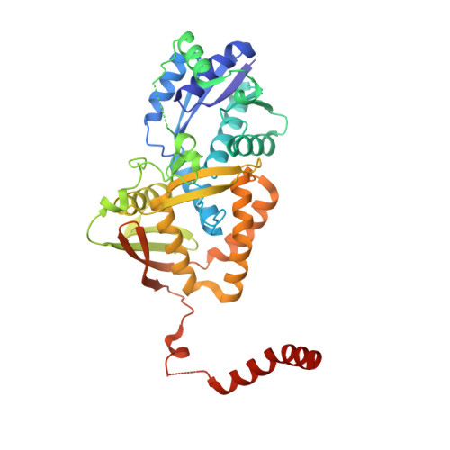
    Argininosuccinate synthetase reversibly catalyzes the ATP-dependent condensation of a citrulline with an aspartate to give argininosuccinate. The structures of the enzyme from Thermus thermophilus HB8 complexed with intact ATP and substrates (citrulline and aspartate) and with AMP and product (argininosuccinate) have been determined at 2.1- and 2.0-A resolution, respectively. The enzyme does not show the ATP-induced domain rotation observed in the enzyme from Escherichia coli. In the enzyme-substrate complex, the reaction sites of ATP and the bound substrates are adjacent and are sufficiently close for the reaction to proceed without the large conformational change at the domain level. The mobility of the triphosphate group in ATP and the side chain of citrulline play an important role in the catalytic action. The protonated amino group of the bound aspartate interacts with the alpha-phosphate of ATP and the ureido group of citrulline, thus stimulating the adenylation of citrulline. The enzyme-product complex explains how the citrullyl-AMP intermediate is bound to the active site. The stereochemistry of the catalysis of the enzyme is clarified on the basis of the structures of tAsS (argininosuccinate synthetase from T. thermophilus HB8) complexes.


  • Organizational Affiliation

    Department of Chemistry, Graduate School of Science, Osaka City University, Osaka 558-8585, Japan.


Macromolecules
Find similar proteins by:  (by identity cutoff)  |  3D Structure
Entity ID: 1
MoleculeChains Sequence LengthOrganismDetailsImage
Argininosuccinate Synthetase
A, B, C, D
400Thermus thermophilusMutation(s): 0 
Gene Names: HB8
EC: 6.3.4.5
UniProt
Find proteins for P59846 (Thermus thermophilus (strain ATCC 27634 / DSM 579 / HB8))
Explore P59846 
Go to UniProtKB:  P59846
Entity Groups  
Sequence Clusters30% Identity50% Identity70% Identity90% Identity95% Identity100% Identity
UniProt GroupP59846
Sequence Annotations
Expand
  • Reference Sequence
Small Molecules
Ligands 3 Unique
IDChains Name / Formula / InChI Key2D Diagram3D Interactions
ATP
Query on ATP

Download Ideal Coordinates CCD File 
E [auth A],
K [auth B],
M [auth C],
O [auth D]
ADENOSINE-5'-TRIPHOSPHATE
C10 H16 N5 O13 P3
ZKHQWZAMYRWXGA-KQYNXXCUSA-N
CIR
Query on CIR

Download Ideal Coordinates CCD File 
F [auth A],
L [auth B],
N [auth C],
P [auth D]
CITRULLINE
C6 H13 N3 O3
RHGKLRLOHDJJDR-BYPYZUCNSA-N
ASP
Query on ASP

Download Ideal Coordinates CCD File 
G [auth A],
H [auth A],
I [auth A],
J [auth A]
ASPARTIC ACID
C4 H7 N O4
CKLJMWTZIZZHCS-REOHCLBHSA-N
Experimental Data & Validation

Experimental Data

  • Method: X-RAY DIFFRACTION
  • Resolution: 2.10 Å
  • R-Value Free: 0.257 
  • R-Value Work: 0.226 
  • Space Group: H 3
Unit Cell:
Length ( Å )Angle ( ˚ )
a = 229.33α = 90
b = 229.33β = 90
c = 159.26γ = 120
Software Package:
Software NamePurpose
HKL-2000data collection
SCALEPACKdata scaling
CNSrefinement
HKL-2000data reduction
CNSphasing

Structure Validation

View Full Validation Report


Entry History 

Deposition Data

Revision History  (Full details and data files)

  • Version 1.0: 2003-04-22
    Type: Initial release
  • Version 1.1: 2008-04-27
    Changes: Version format compliance
  • Version 1.2: 2011-07-13
    Changes: Version format compliance
  • Version 2.0: 2023-11-15
    Changes: Atomic model, Data collection, Database references, Derived calculations