


Crystal Structure of Malate Dehydrogenase from Thermus themrophilus HB8
Hirose, R., Hasegawa, T., Yamano, A., Kuramitsu, S., Hamada, K.To be published.
Experimental Data Snapshot
Starting Model: experimental
View more details
wwPDB Validation 3D Report Full Report

Entity ID: 1 | |||||
|---|---|---|---|---|---|
| Molecule | Chains | Sequence Length | Organism | Details | Image |
| MALATE DEHYDROGENASE | 327 | Thermus thermophilus | Mutation(s): 0 EC: 1.1.1.37 | 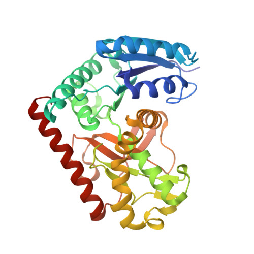 | |
UniProt | |||||
Find proteins for P10584 (Thermus thermophilus) Explore P10584 Go to UniProtKB: P10584 | |||||
Entity Groups | |||||
| Sequence Clusters | 30% Identity50% Identity70% Identity90% Identity95% Identity100% Identity | ||||
| UniProt Group | P10584 | ||||
Sequence AnnotationsExpand | |||||
| |||||
| Length ( Å ) | Angle ( ˚ ) |
|---|---|
| a = 71.64 | α = 90 |
| b = 88.03 | β = 90 |
| c = 118.68 | γ = 90 |
| Software Name | Purpose |
|---|---|
| CrystalClear | data collection |
| CrystalClear | data reduction |
| AMoRE | phasing |
| CNS | refinement |
| CrystalClear | data scaling |
RCSB PDB (citation) is hosted by
RCSB PDB is a member of the

