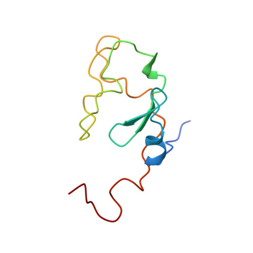
Design challenges for hemoproteins: the solution structure of apocytochrome b5.
Falzone, C.J., Mayer, M.R., Whiteman, E.L., Moore, C.D., Lecomte, J.T.(1996) Biochemistry 35: 6519-6526
- PubMed: 8639599
- DOI: https://doi.org/10.1021/bi960501q
- Primary Citation of Related Structures:
1IET, 1IEU - PubMed Abstract:
In order to characterize the structural and dynamic factors that determine the assembly in b hemoproteins, the solution structure of the 98-residue protein apocytochrome b5 was determined by NMR methods. Over 800 experimental restraints derived from a series of two- and three-dimensional experiments were used. Holocytochrome b5, the protein with iron protoporphyrin-IX liganded to His-39 and His-63, contains in sequence the following elements of secondary structure: beta 1-alpha 1-beta 4-beta 3-alpha 2-alpha 3-beta 5-alpha 4-alpha 5-beta 2-alpha 6 [Mathews, F.S., Czerwinski, E. W., & Argos, P. (1979) The Porphyrins, Vol. 7, pp. 107-147, Academic Press, New York]. The folded holoprotein possesses two hydrophobic cores: an extensive, functional core around the heme (core 1), and a smaller, structural core remote from the heme (core 2). The apoprotein was found to contain a stable four-stranded beta-sheet encompassing beta 1, beta 2, beta 3, and beta 4 and three alpha-helices, corresponding to alpha 1, alpha 2, and alpha 6. Two short alpha-helices (alpha 3 and alpha 5) appear to form partially, and alpha 4 is not detected. These three helices and beta 5 border the heme binding pocket and are disordered in the apoprotein NMR structure. According to backbone 1H-15N NOE results, the most flexible region of the apoprotein, except for the termini, extends from Ala-50 (in beta 5) to Glu-69 (in alpha 5). The polypeptide segment bearing His-63 (located immediately prior to alpha 5) exhibits faster internal motions than that bearing His-39 (at the C-terminal end of alpha 2). The latter imidazole samples a restricted region of space, whereas the former can adopt many orientations with respect to the stable core. It was concluded that heme removal affects the structure and dynamics of most of core 1 whereas it leaves core 2 largely intact. The results provide guidelines for the rational design of b hemoproteins: a modular structure including a packed, stable core and a partially folded binding site is anticipated to present strong kinetic and thermodynamic advantages compared to approaches relying on the complete formation of secondary structure prior to heme binding.
Organizational Affiliation:
Department of Chemistry, Pennsylvania State University, University Park 16802, USA.