1.8 A crystal structure of the C-terminal domain of rabbit serum haemopexin.
Faber, H.R., Groom, C.R., Baker, H.M., Morgan, W.T., Smith, A., Baker, E.N.(1995) Structure 3: 551-559
- PubMed: 8590016
- DOI: https://doi.org/10.1016/s0969-2126(01)00189-7
- Primary Citation of Related Structures:
1HXN - PubMed Abstract:
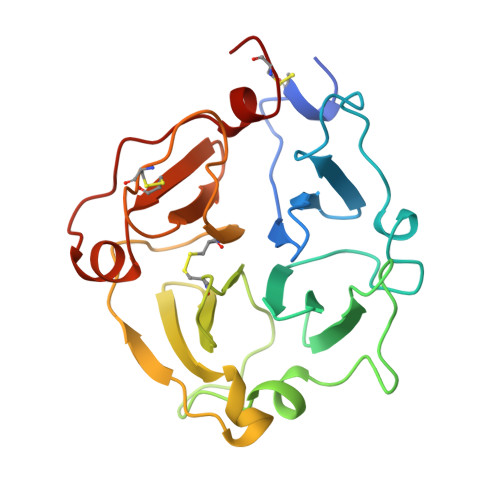
Haemopexin is a serum glycoprotein that binds haem reversibly and delivers it to the liver where it is taken up by receptor-mediated endocytosis. Haemopexin has two homologous domains, each having a characteristic fourfold internal sequence repeat. Haemopexin-type domains are also found in other proteins, including the serum adhesion protein vitronectin and various collagenases, in which they mediate protein-protein interactions. We have determined the crystal structure of the C-terminal domain of haemopexin at 1.8 A resolution. The domain is folded into four beta-leaflet modules, arranged in succession around a central pseudo-fourfold axis. A funnel-shaped tunnel through the centre of this disc-shaped domain serves as an ion-binding site. A model for haem binding by haemopexin is proposed, utilizing an anion-binding site at the wider end of the central tunnel, together with an associated cleft. This parallels the active-site location in other beta-propeller structures. The capacity to bind both cations and anions, together with the disc shape of the domain, suggests that such domains may be used widely for macromolecular recognition.
Organizational Affiliation:
Department of Chemistry and Biochemistry, Massey University, Palmerston North, New Zealand.