1HMV

THE STRUCTURE OF UNLIGANDED REVERSE TRANSCRIPTASE FROM THE HUMAN IMMUNODEFICIENCY VIRUS TYPE 1


Experimental Data Snapshot

  • Method: X-RAY DIFFRACTION
  • Resolution: 3.20 Å
  • R-Value Free: 0.297 
  • R-Value Work: 0.254 
  • R-Value Observed: 0.254 

wwPDB Validation   3D Report Full Report


This is version 1.3 of the entry. See complete history


Literature

The structure of unliganded reverse transcriptase from the human immunodeficiency virus type 1.

Rodgers, D.W.Gamblin, S.J.Harris, B.A.Ray, S.Culp, J.S.Hellmig, B.Woolf, D.J.Debouck, C.Harrison, S.C.

(1995) Proc Natl Acad Sci U S A 92: 1222-1226

  • DOI: https://doi.org/10.1073/pnas.92.4.1222
  • Primary Citation of Related Structures:  
    1HMV

  • PubMed Abstract: 

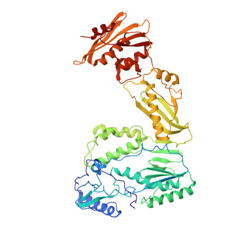
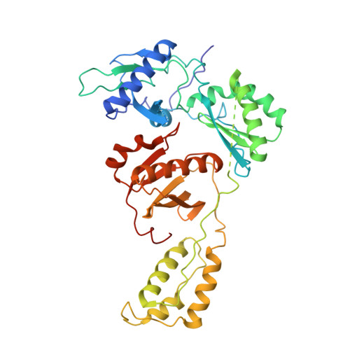
    The crystal structure of the reverse transcriptase (RT) from the type 1 human immunodeficiency virus has been determined at 3.2-A resolution. Comparison with complexes between RT and the polymerase inhibitor Nevirapine [Kohlstaedt, L.A., Wang, J., Friedman, J.M., Rice, P.A. & Steitz, T.A. (1992) Science 256, 1783-1790] and between RT and an oligonucleotide [Jacobo-Molina, A., Ding, J., Nanni, R., Clark, A. D., Lu, X., Tantillo, C., Williams, R. L., Kamer, G., Ferris, A. L., Clark, P., Hizi, A., Hughes, S. H. & Arnold, E. (1993) Proc. Natl. Acad. Sci. USA 90, 6320-6324] reveals changes associated with ligand binding. The enzyme is a heterodimer (p66/p51), with domains labeled "fingers," "thumb," "palm," and "connection" in both subunits, and a ribonuclease H domain in the larger subunit only. The most striking difference between RT and both complex structures is the change in orientation of the p66 thumb (approximately 33 degrees rotation). Smaller shifts relative to the core of the molecule were also found in other domains, including the p66 fingers and palm, which contain the polymerase active site. Within the polymerase catalytic region itself, there are no rearrangements between RT and the RT/DNA complex. In RT/Nevirapine, the drug binds in the p66 palm near the polymerase active site, a region that is well-packed hydrophobic core in the unliganded enzyme. Room for the drug is provided by movement of a small beta-sheet within the palm domain of the Nevirapine complex. The rearrangement within the palm and thumb, as well as domain shifts relative to the enzyme core, may prevent correct placement of the oligonucleotide substrate when the drug is bound.


  • Organizational Affiliation

    Department of Molecular and Cellular Biology, Harvard University, Cambridge, MA 02138.


Macromolecules
Find similar proteins by:  (by identity cutoff)  |  3D Structure
Entity ID: 1
MoleculeChains Sequence LengthOrganismDetailsImage
HIV-1 REVERSE TRANSCRIPTASE (SUBUNIT P66)
A, C, E, G
560Human immunodeficiency virus 1Mutation(s): 0 
Gene Names: HIV-1 POL
EC: 2.7.7.49
UniProt
Find proteins for P03366 (Human immunodeficiency virus type 1 group M subtype B (isolate BH10))
Explore P03366 
Go to UniProtKB:  P03366
Entity Groups  
Sequence Clusters30% Identity50% Identity70% Identity90% Identity95% Identity100% Identity
UniProt GroupP03366
Sequence Annotations
Expand
  • Reference Sequence
Find similar proteins by:  (by identity cutoff)  |  3D Structure
Entity ID: 2
MoleculeChains Sequence LengthOrganismDetailsImage
HIV-1 REVERSE TRANSCRIPTASE (SUBUNIT P51)
B, D, F, H
440Human immunodeficiency virus 1Mutation(s): 0 
Gene Names: HIV-1 POL
EC: 2.7.7.49
UniProt
Find proteins for P03366 (Human immunodeficiency virus type 1 group M subtype B (isolate BH10))
Explore P03366 
Go to UniProtKB:  P03366
Entity Groups  
Sequence Clusters30% Identity50% Identity70% Identity90% Identity95% Identity100% Identity
UniProt GroupP03366
Sequence Annotations
Expand
  • Reference Sequence
Experimental Data & Validation

Experimental Data

  • Method: X-RAY DIFFRACTION
  • Resolution: 3.20 Å
  • R-Value Free: 0.297 
  • R-Value Work: 0.254 
  • R-Value Observed: 0.254 
  • Space Group: C 1 2 1
Unit Cell:
Length ( Å )Angle ( ˚ )
a = 168.7α = 90
b = 162.8β = 105.7
c = 331.8γ = 90
Software Package:
Software NamePurpose
X-PLORmodel building
X-PLORrefinement
X-PLORphasing

Structure Validation

View Full Validation Report


Entry History 

Deposition Data

Revision History  (Full details and data files)

  • Version 1.0: 1995-03-31
    Type: Initial release
  • Version 1.1: 2008-03-24
    Changes: Version format compliance
  • Version 1.2: 2011-07-13
    Changes: Version format compliance
  • Version 1.3: 2024-02-07
    Changes: Data collection, Database references, Derived calculations, Other