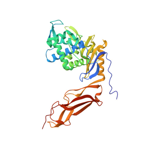
Crystal Structure of a Deacylation-Defective Mutant of Penicillin-Binding Protein 5 at 2.3-A Resolution
Davies, C., White, S.W., Nicholas, R.A.(2001) J Biol Chem 276: 616
- PubMed: 10967102
- DOI: https://doi.org/10.1074/jbc.M004471200
- Primary Citation of Related Structures:
1HD8 - PubMed Abstract:
Penicillin-binding protein 5 (PBP 5) of Escherichia coli functions as a d-alanine carboxypeptidase, cleaving the C-terminal d-alanine residue from cell wall peptides. Like all PBPs, PBP 5 forms a covalent acyl-enzyme complex with beta-lactam antibiotics; however, PBP 5 is distinguished by its high rate of deacylation of the acyl-enzyme complex (t(12) approximately 9 min). A Gly-105 --> Asp mutation in PBP 5 markedly impairs this beta-lactamase activity (deacylation), with only minor effects on acylation, and promotes accumulation of a covalent complex with peptide substrates. To gain further insight into the catalytic mechanism of PBP 5, we determined the three-dimensional structure of the G105D mutant form of soluble PBP 5 (termed sPBP 5') at 2.3 A resolution. The structure is composed of two domains, a penicillin binding domain with a striking similarity to Class A beta-lactamases (TEM-1-like) and a domain of unknown function. In addition, the penicillin-binding domain contains an active site loop spatially equivalent to the Omega loop of beta-lactamases. In beta-lactamases, the Omega loop contains two amino acids involved in catalyzing deacylation. This similarity may explain the high beta-lactamase activity of wild-type PBP 5. Because of the low rate of deacylation of the G105D mutant, visualization of peptide substrates bound to the active site may be possible.
Organizational Affiliation:
School of Biological Sciences, University of Sussex, Falmer, Brighton BN1 9QG, United Kingdom.