Folded-Back Solution Structure of Monomeric Factor H of Human Complement by Synchrotron X-Ray and Neutron Scattering, Analytical Ultracentrifugation and Constrained Molecular Modelling.
Aslam, M., Perkins, S.J.(2001) J Mol Biol 309: 1117
- PubMed: 11399083
- DOI: https://doi.org/10.1006/jmbi.2001.4720
- Primary Citation of Related Structures:
1HAQ - PubMed Abstract:
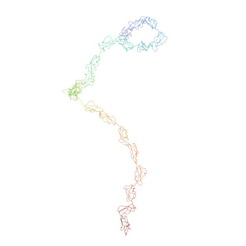
Factor H (FH) is a regulatory cofactor for the protease factor I in the breakdown of C3b in the complement system of immune defence, and binds to heparin and other polyanionic substrates. FH is composed of 20 short consensus/complement repeat (SCR) domains, for which the overall arrangement in solution is unknown. As previous studies had shown that FH can form monomeric or dimeric structures, X-ray and neutron scattering was accordingly performed with FH in the concentration range between 0.7 and 14 mg ml(-1). The radius of gyration of FH was determined to be 11.1-11.3 nm by both methods, and the radii of gyration of the cross-section were 4.4 nm and 1.7 nm. The distance distribution function P(r) showed that the overall length of FH was 38 nm. The neutron data showed that FH was monomeric with a molecular mass of 165,000(+/-17,000) Da. Analytical ultracentrifugation data confirmed this, where sedimentation equilibrium curve fits gave a mean molecular mass of 155,000(+/-3,000) Da. Sedimentation velocity experiments using the g*(s) derivative method showed that FH was monodisperse and had a sedimentation coefficient of 5.3(+/-0.1) S. In order to construct a full model of FH for scattering curve and sedimentation coefficient fits, homology models were constructed for 17 of the 20 SCR domains using knowledge of the NMR structures for FH SCR-5, SCR-15 and SCR-16, and vaccinia coat protein SCR-3 and SCR-4. Molecular dynamics simulations were used to generate a large conformational library for each of the 19 SCR-SCR linker peptides. Peptides from these libraries were combined with the 20 SCR structures in order to generate stereochemically complete models for the FH structure. Using an automated constrained fit procedure, the analysis of 16,752 possible FH models showed that only those models in which the 20 SCR domains were bent back upon themselves were able to account for the scattering and sedimentation data. The best-fit models showed that FH had an overall length of 38 nm and is flexible. This length is significantly less than a predicted length of 73 nm if the 20 SCR structures had been arranged in an extended arrangement. This outcome is attributed to several long linker sequences. These bent-back domain structures may correspond to conformational flexibility in FH and enable the multiple FH binding sites for C3 and heparin to come into close proximity.
Organizational Affiliation:
Department of Biochemistry and Molecular Biology, Royal Free Campus, Royal Free and University College Medical School, University College London, Rowland Hill Street, London, NW3 2PF, UK.