1GR0

myo-inositol 1-phosphate synthase from Mycobacterium tuberculosis in complex with NAD and zinc.


Experimental Data Snapshot

  • Method: X-RAY DIFFRACTION
  • Resolution: 1.95 Å
  • R-Value Free: 0.239 
  • R-Value Work: 0.211 
  • R-Value Observed: 0.211 

wwPDB Validation   3D Report Full Report


This is version 1.3 of the entry. See complete history


Literature

Crystal Structure of Inositol 1-Phosphate Synthase from Mycobacterium Tuberculosis, a Key Enzyme in Phosphatidylinositol Synthesis

Norman, R.A.Mcalister, M.S.B.Murray-Rust, J.Movahedzadeh, F.Stoker, N.G.Mcdonald, N.Q.

(2002) Structure 10: 393

  • DOI: https://doi.org/10.1016/s0969-2126(02)00718-9
  • Primary Citation of Related Structures:  
    1GR0

  • PubMed Abstract: 

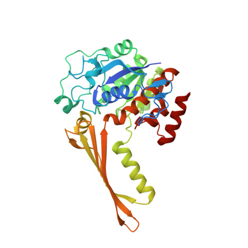
    Phosphatidylinositol (PI) is essential for Mycobacterium tuberculosis viability and the enzymes involved in the PI biosynthetic pathway are potential antimycobacterial agents for which little structural information is available. The rate-limiting step in the pathway is the production of (L)-myo-inositol 1-phosphate from (D)-glucose 6-phosphate, a complex reaction catalyzed by the enzyme inositol 1-phosphate synthase. We have determined the crystal structure of this enzyme from Mycobacterium tuberculosis (tbINO) at 1.95 A resolution, bound to the cofactor NAD+. The active site is located within a deep cleft at the junction between two domains. The unexpected presence of a zinc ion here suggests a mechanistic difference from the eukaryotic inositol synthases, which are stimulated by monovalent cations, that may be exploitable in developing selective inhibitors of tbINO.


  • Organizational Affiliation

    Structural Biology Laboratory, Cancer Research U K, London, United Kingdom.


Macromolecules
Find similar proteins by:  (by identity cutoff)  |  3D Structure
Entity ID: 1
MoleculeChains Sequence LengthOrganismDetailsImage
INOSITOL-3-PHOSPHATE SYNTHASE367Mycobacterium tuberculosisMutation(s): 0 
EC: 5.5.1.4
UniProt
Find proteins for P9WKI1 (Mycobacterium tuberculosis (strain ATCC 25618 / H37Rv))
Explore P9WKI1 
Go to UniProtKB:  P9WKI1
Entity Groups  
Sequence Clusters30% Identity50% Identity70% Identity90% Identity95% Identity100% Identity
UniProt GroupP9WKI1
Sequence Annotations
Expand
  • Reference Sequence
Small Molecules
Ligands 3 Unique
IDChains Name / Formula / InChI Key2D Diagram3D Interactions
NAD
Query on NAD

Download Ideal Coordinates CCD File 
B [auth A]NICOTINAMIDE-ADENINE-DINUCLEOTIDE
C21 H27 N7 O14 P2
BAWFJGJZGIEFAR-NNYOXOHSSA-N
CAC
Query on CAC

Download Ideal Coordinates CCD File 
D [auth A]CACODYLATE ION
C2 H6 As O2
OGGXGZAMXPVRFZ-UHFFFAOYSA-M
ZN
Query on ZN

Download Ideal Coordinates CCD File 
C [auth A]ZINC ION
Zn
PTFCDOFLOPIGGS-UHFFFAOYSA-N
Experimental Data & Validation

Experimental Data

  • Method: X-RAY DIFFRACTION
  • Resolution: 1.95 Å
  • R-Value Free: 0.239 
  • R-Value Work: 0.211 
  • R-Value Observed: 0.211 
  • Space Group: P 4 21 2
Unit Cell:
Length ( Å )Angle ( ˚ )
a = 116.195α = 90
b = 116.195β = 90
c = 64.544γ = 90
Software Package:
Software NamePurpose
CNSrefinement
DENZOdata reduction
SCALEPACKdata scaling
CNSphasing

Structure Validation

View Full Validation Report


Entry History 

Deposition Data

Revision History  (Full details and data files)

  • Version 1.0: 2002-03-12
    Type: Initial release
  • Version 1.1: 2011-09-07
    Changes: Derived calculations, Non-polymer description, Other, Refinement description, Structure summary, Version format compliance
  • Version 1.2: 2016-12-21
    Changes: Structure summary
  • Version 1.3: 2019-08-21
    Changes: Data collection, Database references