1FZE

CRYSTAL STRUCTURE OF FRAGMENT DOUBLE-D FROM HUMAN FIBRIN


Experimental Data Snapshot

  • Method: X-RAY DIFFRACTION
  • Resolution: 3.00 Å
  • R-Value Free: 0.318 
  • R-Value Work: 0.251 
  • R-Value Observed: 0.251 

wwPDB Validation   3D Report Full Report


This is version 1.4 of the entry. See complete history


Literature

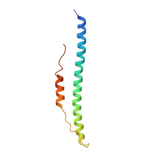
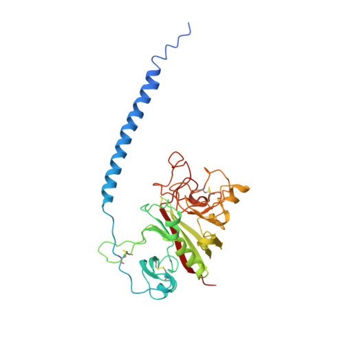
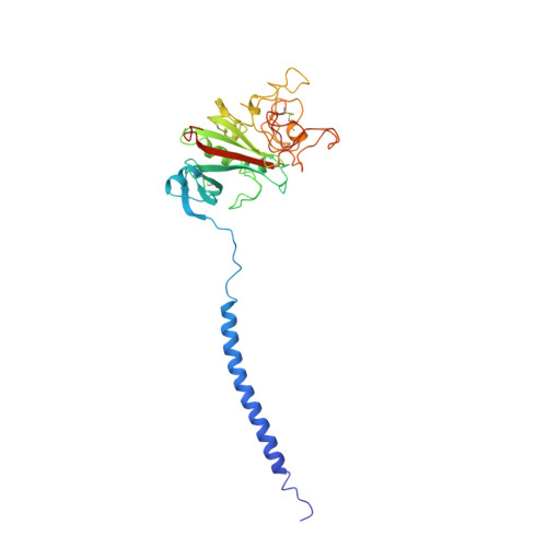
Conformational changes in fragments D and double-D from human fibrin(ogen) upon binding the peptide ligand Gly-His-Arg-Pro-amide.

Everse, S.J.Spraggon, G.Veerapandian, L.Doolittle, R.F.

(1999) Biochemistry 38: 2941-2946

  • DOI: https://doi.org/10.1021/bi982626w
  • Primary Citation of Related Structures:  
    1FZE, 1FZF, 1FZG

  • PubMed Abstract: 

    The structure of fragment double-D from human fibrin has been solved in the presence and absence of the peptide ligands that simulate the two knobs exposed by the removal of fibrinopeptides A and B, respectively. All told, six crystal structures have been determined, three of which are reported here for the first time: namely, fragments D and double-D with the peptide GHRPam alone and double-D in the absence of any peptide ligand. Comparison of the structures has revealed a series of conformational changes that are brought about by the various knob-hole interactions. Of greatest interest is a moveable "flap" of two negatively charged amino acids (Glubeta397 and Aspbeta398) whose side chains are pinned back to the coiled coil with a calcium atom bridge until GHRPam occupies the beta-chain pocket. Additionally, in the absence of the peptide ligand GPRPam, GHRPam binds to the gamma-chain pocket, a new calcium-binding site being formed concomitantly.


  • Organizational Affiliation

    Center for Molecular Genetics, University of California, San Diego, La Jolla 92093-0634, USA.


Macromolecules
Find similar proteins by:  (by identity cutoff)  |  3D Structure
Entity ID: 1
MoleculeChains Sequence LengthOrganismDetailsImage
FIBRINOGEN
A, D
87Homo sapiensMutation(s): 0 
UniProt & NIH Common Fund Data Resources
Find proteins for P02671 (Homo sapiens)
Explore P02671 
Go to UniProtKB:  P02671
PHAROS:  P02671
GTEx:  ENSG00000171560 
Entity Groups  
Sequence Clusters30% Identity50% Identity70% Identity90% Identity95% Identity100% Identity
UniProt GroupP02671
Sequence Annotations
Expand
  • Reference Sequence
Find similar proteins by:  (by identity cutoff)  |  3D Structure
Entity ID: 2
MoleculeChains Sequence LengthOrganismDetailsImage
FIBRINOGEN
B, E
328Homo sapiensMutation(s): 0 
UniProt & NIH Common Fund Data Resources
Find proteins for P02675 (Homo sapiens)
Explore P02675 
Go to UniProtKB:  P02675
PHAROS:  P02675
GTEx:  ENSG00000171564 
Entity Groups  
Sequence Clusters30% Identity50% Identity70% Identity90% Identity95% Identity100% Identity
UniProt GroupP02675
Sequence Annotations
Expand
  • Reference Sequence
Find similar proteins by:  (by identity cutoff)  |  3D Structure
Entity ID: 3
MoleculeChains Sequence LengthOrganismDetailsImage
FIBRINOGEN
C, F
319Homo sapiensMutation(s): 0 
UniProt & NIH Common Fund Data Resources
Find proteins for P02679 (Homo sapiens)
Explore P02679 
Go to UniProtKB:  P02679
PHAROS:  P02679
GTEx:  ENSG00000171557 
Entity Groups  
Sequence Clusters30% Identity50% Identity70% Identity90% Identity95% Identity100% Identity
UniProt GroupP02679
Sequence Annotations
Expand
  • Reference Sequence
Experimental Data & Validation

Experimental Data

  • Method: X-RAY DIFFRACTION
  • Resolution: 3.00 Å
  • R-Value Free: 0.318 
  • R-Value Work: 0.251 
  • R-Value Observed: 0.251 
  • Space Group: P 1 21 1
Unit Cell:
Length ( Å )Angle ( ˚ )
a = 108α = 90
b = 48.6β = 104.4
c = 166.4γ = 90
Software Package:
Software NamePurpose
DENZOdata reduction
SCALEPACKdata scaling
X-PLORmodel building
X-PLORrefinement
X-PLORphasing

Structure Validation

View Full Validation Report


Entry History 

Deposition Data

Revision History  (Full details and data files)

  • Version 1.0: 1999-06-08
    Type: Initial release
  • Version 1.1: 2008-03-24
    Changes: Version format compliance
  • Version 1.2: 2011-07-13
    Changes: Derived calculations, Version format compliance
  • Version 1.3: 2020-07-29
    Type: Remediation
    Reason: Carbohydrate remediation
    Changes: Advisory, Data collection, Derived calculations, Structure summary
  • Version 1.4: 2023-08-09
    Changes: Database references, Refinement description, Structure summary