1FQX

CRYSTAL STRUCTURE OF THE COMPLEX OF HIV-1 PROTEASE WITH A PEPTIDOMIMETIC INHIBITOR


Experimental Data Snapshot

  • Method: X-RAY DIFFRACTION
  • Resolution: 3.10 Å
  • R-Value Work: 0.180 
  • R-Value Observed: 0.180 

wwPDB Validation   3D Report Full Report


Ligand Structure Quality Assessment 


This is version 1.4 of the entry. See complete history


Literature

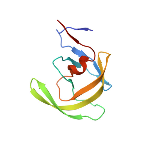
A distinct binding mode of a hydroxyethylamine isostere inhibitor of HIV-1 protease.

Dohnalek, J.Hasek, J.Duskova, J.Petrokova, H.Hradilek, M.Soucek, M.Konvalinka, J.Brynda, J.Sedlacek, J.Fabry, M.

(2001) Acta Crystallogr D Biol Crystallogr 57: 472-476

  • DOI: https://doi.org/10.1107/s0907444900018928
  • Primary Citation of Related Structures:  
    1FQX

  • PubMed Abstract: 

    Crystallization conditions for an HIV-1 protease-inhibitor complex were optimized to produce crystals suitable for X-ray diffraction experiments. The X-ray structure of the HIV-1 protease complex was solved and refined at 3.1 A resolution. In contrast to Saquinavir, the mimetic hydroxy group of the inhibitor Boc-Phe-Psi[(S)-CH(OH)CH(2)NH]-Phe-Glu-Phe-NH(2) is placed asymmetrically with respect to the non-crystallographic twofold axis of the protease dimer so that hydrogen bonds between the amino group of the inhibitor and the catalytic aspartates can be formed. The inhibitor binds in the centre of the active site by a compact network of hydrogen bonds to Gly27, Gly127, Asp25, Asp125 and via the buried water molecule W301 to Ile50 and Ile150.


  • Organizational Affiliation

    Institute of Macromolecular Chemistry, Academy of Sciences of the Czech Republic, Heyrovského nám. 2, 162 06 Praha 6, Czech Republic. dohnalek@imc.cas.cz


Macromolecules
Find similar proteins by:  (by identity cutoff)  |  3D Structure
Entity ID: 1
MoleculeChains Sequence LengthOrganismDetailsImage
PROTEASE RETROPEPSIN
A, B
99Human immunodeficiency virus 1Mutation(s): 0 
EC: 3.4.23.16
UniProt
Find proteins for P03367 (Human immunodeficiency virus type 1 group M subtype B (isolate BRU/LAI))
Explore P03367 
Go to UniProtKB:  P03367
Entity Groups  
Sequence Clusters30% Identity50% Identity70% Identity90% Identity95% Identity100% Identity
UniProt GroupP03367
Sequence Annotations
Expand
  • Reference Sequence
Small Molecules
Ligands 1 Unique
IDChains Name / Formula / InChI Key2D Diagram3D Interactions
0ZT
Query on 0ZT

Download Ideal Coordinates CCD File 
C [auth A]N-{(2S,3S)-3-[(tert-butoxycarbonyl)amino]-2-hydroxy-4-phenylbutyl}-L-phenylalanyl-L-alpha-glutamyl-L-phenylalaninamide
C38 H49 N5 O8
MPMUDVMRFYJRLP-XDIGFQIYSA-N
Binding Affinity Annotations 
IDSourceBinding Affinity
0ZT BindingDB:  1FQX Ki: min: 0.15, max: 86 (nM) from 3 assay(s)
Biologically Interesting Molecules (External Reference) 1 Unique
Experimental Data & Validation

Experimental Data

Unit Cell:
Length ( Å )Angle ( ˚ )
a = 63.13α = 90
b = 63.13β = 90
c = 83.12γ = 120
Software Package:
Software NamePurpose
AMoREphasing
CNSrefinement
DENZOdata reduction
SCALEPACKdata scaling

Structure Validation

View Full Validation Report


Ligand Structure Quality Assessment 


Entry History 

Deposition Data

Revision History  (Full details and data files)

  • Version 1.0: 2001-03-14
    Type: Initial release
  • Version 1.1: 2007-10-16
    Changes: Version format compliance
  • Version 1.2: 2011-07-13
    Changes: Atomic model, Database references, Derived calculations, Non-polymer description, Structure summary, Version format compliance
  • Version 1.3: 2012-12-12
    Changes: Other
  • Version 1.4: 2024-02-07
    Changes: Data collection, Database references, Derived calculations