Crystal structure of a 30 kDa C-terminal fragment from the gamma chain of human fibrinogen.
Yee, V.C., Pratt, K.P., Cote, H.C., Trong, I.L., Chung, D.W., Davie, E.W., Stenkamp, R.E., Teller, D.C.(1997) Structure 5: 125-138
- PubMed: 9016719
- DOI: https://doi.org/10.1016/s0969-2126(97)00171-8
- Primary Citation of Related Structures:
1FIB, 1FIC, 1FID - PubMed Abstract:
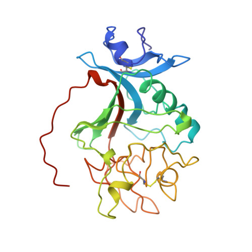
Blood coagulation occurs by a cascade of zymogen activation resulting from minor proteolysis. The final stage of coagulation involves thrombin generation and limited proteolysis of fibrinogen to give spontaneously polymerizing fibrin. The resulting fibrin network is covalently crosslinked by factor XIIIa to yield a stable blood clot. Fibrinogen is a 340 kDa glycoprotein composed of six polypeptide chains, (alphabetagamma)2, held together by 29 disulfide bonds. The globular C terminus of the gamma chain contains a fibrin-polymerization surface, the principal factor XIIIa crosslinking site, the platelet receptor recognition site, and a calcium-binding site. Structural information on this domain should thus prove helpful in understanding clot formation. The X-ray crystallographic structure of the 30 kDa globular C terminus of the gamma chain of human fibrinogen has been determined in one crystal form using multiple isomorphous replacement methods. The refined coordinates were used to solve the structure in two more crystal forms by molecular replacement; the crystal structures have been refined against diffraction data to either 2.5 A or 2.1 A resolution. Three domains were identified in the structure, including a C-terminal fibrin-polymerization domain (P), which contains a single calcium-binding site and a deep binding pocket that provides the polymerization surface. The overall structure has a pronounced dipole moment, and the C-terminal residues appear highly flexible. The polymerization domain in the gamma chain is the most variable among a family of fibrinogen-related proteins and contains many acidic residues. These residues contribute to the molecular dipole moment in the structure, which may allow electrostatic steering to guide the alignment of fibrin monomers during the polymerization process. The flexibility of the C-terminal residues, which contain one of the factor XIIIa crosslinking sites and the platelet receptor recognition site, may be important in the function of this domain.
Organizational Affiliation:
Department of Biochemistry, Biomolecular Structure Center, University of Washington, Seattle, WA 98195, USA.