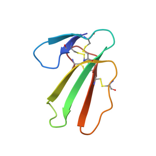
Membrane binding motif of the P-type cardiotoxin.
Dubovskii, P.V., Dementieva, D.V., Bocharov, E.V., Utkin, Y.N., Arseniev, A.S.(2001) J Mol Biol 305: 137-149
- PubMed: 11114253
- DOI: https://doi.org/10.1006/jmbi.2000.4283
- Primary Citation of Related Structures:
1FFJ - PubMed Abstract:
Carditoxins (CTXs) from cobra snake venoms, the basic 60-62 residue all-beta sheet polypeptides, are known to bind to and impair the function of cell membranes. To assess the membrane induced conformation and orientation of CTXs, the interaction of the P-type cardiotoxin II from Naja oxiana snake venom (CTII) with perdeuterated dodecylphosphocholine (DPC) was studied using ( 1 )H-NMR spectroscopy and diffusion measurements. Under conditions where the toxin formed a well-defined complex with DPC, the spatial structure of CTII with respect to the presence of tightly bound water molecules in loop II, was calculated using the torsion angle dynamics program DYANA. The structure was found to be similar, except for subtle changes in the tips of all three loops, to the previously described "major" form of CTII in aqueous solution illustrated by the "trans" configuration of the Val7-Pro8 peptide bond. No "minor" form with the "cis" configuration of the above bond was found in the micelle-bound state. The broadening of the CTII backbone proton signals by 5, 16-doxylstearate relaxation probes, together with modeling based on the spatial structure of CTII, indicated a periphery mode of binding of the toxin molecule to the micelle and revealed its micelle interacting domain. The latter includes a hydrophobic region of CTII within the extremities of loops I and III (residues 5-11, 46-50), the basement of loop II (residues 24-29,31-37) and the belt of polar residues encircling these loops (lysines 4,5,12,23,50, serines 11,46, histidine 31, arginine 36). It is suggested that this structural motif and the mode of binding can be realized during interaction of CTXs with lipid and biological membranes.
Organizational Affiliation:
Shemyakin & Ovchinnikov Institute of Bioorganic Chemistry, Russian Academy of Sciences, 16/10 Miklukho-Maklaya str., V-437, Moscow, Russia.