1FFF

STRUCTURAL IMPLICATIONS OF DRUG RESISTANT MUTANTS OF HIV-1 PROTEASE : HIGH RESOLUTION CRYSTAL STRUCTURES OF THE MUTANT PROTEASE/SUBSTRATE ANALOG COMPLEXES.


Experimental Data Snapshot

  • Method: X-RAY DIFFRACTION
  • Resolution: 1.90 Å
  • R-Value Free: 0.248 
  • R-Value Work: 0.212 

wwPDB Validation   3D Report Full Report


This is version 1.6 of the entry. See complete history


Literature

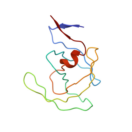
Structural implications of drug-resistant mutants of HIV-1 protease: high-resolution crystal structures of the mutant protease/substrate analogue complexes.

Mahalingam, B.Louis, J.M.Hung, J.Harrison, R.W.Weber, I.T.

(2001) Proteins 43: 455-464

  • DOI: https://doi.org/10.1002/prot.1057
  • Primary Citation of Related Structures:  
    1FEJ, 1FF0, 1FFF, 1FFI, 1FG6, 1FG8, 1FGC

  • PubMed Abstract: 

    Emergence of drug-resistant mutants of HIV-1 protease is an ongoing problem in the fight against AIDS. The mechanisms governing resistance are both complex and varied. We have determined crystal structures of HIV-1 protease mutants, D30N, K45I, N88D, and L90M complexed with peptide inhibitor analogues of CA-p2 and p2-NC cleavage sites in the Gag-pol precursor in order to study the structural mechanisms underlying resistance. The structures were determined at 1.55-1.9-A resolution and compared with the wild-type structure. The conformational disorder seen for most of the hydrophobic side-chains around the inhibitor binding site indicates flexibility of binding. Eight water molecules are conserved in all 9 structures; their location suggests that they are important for catalysis as well as structural stability. Structural differences among the mutants were analyzed in relation to the observed changes in protease activity and stability. Mutant L90M shows steric contacts with the catalytic Asp25 that could destabilize the catalytic loop at the dimer interface, leading to its observed decreased dimer stability and activity. Mutant K45I reduces the mobility of the flap and the inhibitor and contributes to an enhancement in structural stability and activity. The side-chain variations at residue 30 relative to wild-type are the largest in D30N and the changes are consistent with the altered activity observed with peptide substrates. Polar interactions in D30N are maintained, in agreement with the observed urea sensitivity. The side-chains of D30N and N88D are linked through a water molecule suggesting correlated changes at the two sites, as seen with clinical inhibitors. Structural changes seen in N88D are small; however, water molecules that mediate interactions between Asn88 and Thr74/Thr31/Asp30 in other complexes are missing in N88D.


  • Organizational Affiliation

    Department of Biology, Georgia State University, Atlanta, Georgia 30303, USA.


Macromolecules
Find similar proteins by:  (by identity cutoff)  |  3D Structure
Entity ID: 1
MoleculeChains Sequence LengthOrganismDetailsImage
PROTEASE RETROPEPSINA [auth C],
B [auth D]
99Human immunodeficiency virus 1Mutation(s): 6 
EC: 3.4.23.16
UniProt
Find proteins for P04587 (Human immunodeficiency virus type 1 group M subtype B (isolate BH5))
Explore P04587 
Go to UniProtKB:  P04587
Entity Groups  
Sequence Clusters30% Identity50% Identity70% Identity90% Identity95% Identity100% Identity
UniProt GroupP04587
Sequence Annotations
Expand
  • Reference Sequence
Small Molecules
Ligands 1 Unique
IDChains Name / Formula / InChI Key2D Diagram3D Interactions
0Q4
Query on 0Q4

Download Ideal Coordinates CCD File 
C [auth D]N-[(2R)-2-({N~5~-[amino(iminio)methyl]-L-ornithyl-L-valyl}amino)-4-methylpentyl]-L-phenylalanyl-L-alpha-glutamyl-L-alanyl-L-norleucinamide
C40 H70 N11 O8
SGWAZUZKMXHYMB-UQGDEETHSA-O
Experimental Data & Validation

Experimental Data

  • Method: X-RAY DIFFRACTION
  • Resolution: 1.90 Å
  • R-Value Free: 0.248 
  • R-Value Work: 0.212 
  • Space Group: P 21 21 21
Unit Cell:
Length ( Å )Angle ( ˚ )
a = 50.908α = 90
b = 58.255β = 90
c = 61.148γ = 90
Software Package:
Software NamePurpose
AMoREphasing
X-PLORrefinement
ADSCdata collection
DENZOdata reduction
SCALEPACKdata scaling

Structure Validation

View Full Validation Report


Entry History 

Deposition Data

Revision History  (Full details and data files)

  • Version 1.0: 2001-06-01
    Type: Initial release
  • Version 1.1: 2008-04-27
    Changes: Version format compliance
  • Version 1.2: 2011-07-13
    Changes: Atomic model, Database references, Derived calculations, Non-polymer description, Structure summary, Version format compliance
  • Version 1.3: 2012-12-12
    Changes: Other
  • Version 1.4: 2021-11-03
    Changes: Database references, Derived calculations
  • Version 1.5: 2024-02-07
    Changes: Data collection
  • Version 1.6: 2024-03-13
    Changes: Source and taxonomy, Structure summary