Hydration, mobility and accessibility of lysozyme: structures of a pH 6.5 orthorhombic form and its low-humidity variant and a comparative study involving 20 crystallographically independent molecules.
Biswal, B.K., Sukumar, N., Vijayan, M.(2000) Acta Crystallogr D Biol Crystallogr 56: 1110-1119
- PubMed: 10957630
- DOI: https://doi.org/10.1107/s0907444900008866
- Primary Citation of Related Structures:
1F0W, 1F10 - PubMed Abstract:
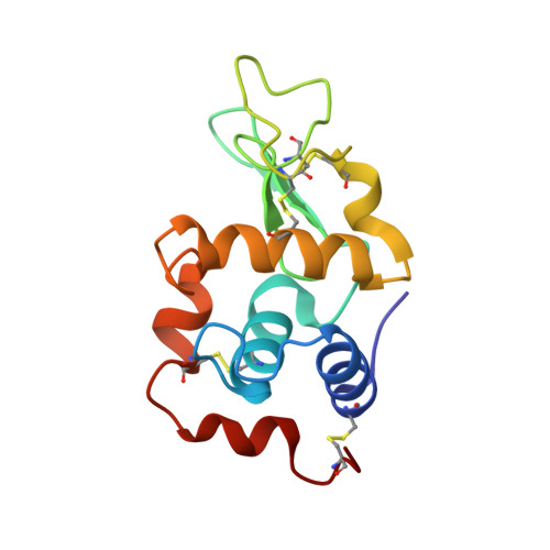
The structure analyses of orthorhombic lysozyme grown at pH 6.5 and its low-humidity variant are reported. The structures of the same form grown at pH 9.5 and 4.5 and that of the low-humidity variant of the pH 9.5 form are available. A comparison between them shows that the changes in molecular geometry and hydration caused by changes in the amount of solvent surrounding protein molecules are more pronounced than those caused by variation in pH. In particular, the conformation and the mutual orientation of the catalytic residues Glu35 and Asp52 remain unaffected by change in pH. A comparative study involving 20 crystallographically independent lysozyme molecules, including five in the orthorhombic form, leads to the delineation of the relatively rigid, moderately flexible and highly flexible regions of the molecule. Half the binding cleft (subsites D, E and F) belong to the rigid region but the other half (subsites A, B and C) belong to a flexible region. There is no marked correlation between relative rigidity and conservation of side-chain conformation except at the binding site. The study permits the identification of seven invariant water molecules associated with the protein. Most of them are involved in important tertiary interactions, while one occurs in the active-site cleft. The study demonstrates a weak correlation between non-accessibility and rigidity. On average, the level of hydration of polar atoms increases rapidly with accessible atomic surface area, but levels off at about 15 A(2) at a little over one ordered water molecule per polar protein atom. Only 15 N and O atoms are hydrated in all 20 molecules. 13 of these are hydrated by the seven invariant water molecules. Of the seven, only one water molecule is totally buried within the protein.
Organizational Affiliation:
Molecular Biophysics Unit, Indian Institute of Science, Bangalore 560 012, India.